
庄文颖 , 曾昭清, 刘晓夏
中国科学院微生物研究所真菌学国家重点实验室 北京 100101
ZHUANG Wen-Ying, ZENG Zhao-Qing, LIU Xiao-Xia
展开
摘要
提供了中国二头孢盘菌属的分类研究概况和已知种类。对属的概念进行了修订;基于形态学特征和序列分析的结果,将兰斯盘菌属的3个种转入二头孢盘菌属,建立3个新组合(黄二头孢盘菌、黄山二头孢盘菌、暗丝二头孢盘菌);描述了一新种(缩孢二头孢盘菌)。该属目前已知的10个种中,在我国分布9个种。编制了该属世界已知种的分种检索表。
关键词:
Abstract
Studies of the genus Dicephalospora from China are briefly reviewed. The generic concept of Dicephalospora is emended. Three new combinations are established based on morphology and ITS sequence data for three taxa previously assigned to the genus Lanzia. A new species is described. Among the ten currently known species in the world, nine of them are distributed in China. A key to the known species of the genus is provided.
Keywords:
The genus Dicephalospora Spooner was established originally for two inoperculate cup-fungi on stromatized plant tissues, which have ectal excipulum of textura prismatica, ectal excipular cell walls slightly thickened and refractive, the poles of ascospores capped with a mucilaginous collar, and asci J+ in Melzer’s reagent (Spooner 1987). In the Flora Fungorum Sinicorum Vol. 8 Sclerotiniaceae et Geoglossaceae (Zhuang 1998), a single species Dicephalospora rufocornea was recorded from China, which is a widely distributed fungus not only in China but also in other regions of the world (Dumont 1980; Spooner 1987). The genus was thought to be morphologically similar to Lanzia Sacc. and is distinguishable and diagnostic by the presence of a hyaline gel collar at each end of ascospores.
Later investigations revealed that China has a relatively high species diversity of Dicephalospora, and three more species were discovered, i.e. D. calochroa (Syd.) Spooner (Wu et al. 1996), D. damingshanica W.Y. Zhuang (Zhuang 1999) and D. pinglongshanica W.Y. Zhuang (Zhuang 1999). At the time, the country has almost all the world known species of the genus. Recently, Dicephalospora chrysotricha (Berk.) Verkley was added to the group (Verkley 2004), which has not been found in China. And Dicephalospora dentata Xiao X. Liu & W.Y. Zhuang characterized by the apothecia with a dentate margin is just described from tropical China (Liu et al. 2015).
In the previous sequence analyses of selected genera of inoperculate cup-fungi (Zhuang & Liu 2007; Liu et al. 2015), we noticed that Lanzia huangshanica W.Y. Zhuang and L. aurantiaca (W.Y. Zhuang) W.Y. Zhuang are closely related to Dicephalospora rufocornea other than members of Lanzia which might not be a monophyletic genus. However, formal nomenclatural changes were not being made. In this study, we emend the generic concept of Dicephalospora based on morphology and sequence analyses of internal transcribed spacer (ITS), and transfer three species from Lanzia to Dicephalospora. Up to now, ten species of the genus are known in the world, and nine of them occur in China.
Specimens of Dicephalospora examined were deposited in the Herbarium Mycologicum Academiae Sinicae (HMAS). Dried apothecia were rehydrated in distilled water and sectioned with a freezing microtome (YD-1508-III, Yidi Medical Appliance Factory, Jinhua, Zhejiang) at a thickness of 15μm. Photographs were taken using a Canon G5 digital camera (Tokyo, Japan) connected to a Zeiss Axioskop 2 Plus microscope (Göttingen, Germany) for anatomical structures and to a Zeiss Stemi 2000C stereomicroscope for gross morphology.
DNA was extracted from dried apothecia or mycelium harvested from colonies on PDA following the methods of Wang & Zhuang (2004). The ITS region was amplified and sequenced with the primer pairs ITS5 and ITS4 (White et al. 1990). PCR was performed with the 2720 Thermal cycler (Applied Biosystems, Foster City, California, USA) with a 25μL reaction system consisting of 12.5μL Taq MasterMix, 1μL each primer (10μmol/L), 1μL template DNA and 9.5μL ddH2O. PCR conditions were an initial step of 5min at 94°C, 30 cycles of 30s at 94°C, 30s at 53°C, and 30s at 72°C, followed by 10min at 72°C. DNA sequencing was carried out in both directions with an ABI 3730 XL DNA Sequencer (SinoGenoMax Co. Ltd.). Sequences achieved in this study were submitted to GenBank (Table 1).
DNA sequences were aligned using ClustalX 1.8 (Thompson et al. 1997), and the alignments were visually adjusted by BioEdit 7.0.5 (Hall 1999). The neighbor-joining (NJ) analysis was carried out using PAUP*4.0b10 (Swofford 2002) with 1 000 bootstrap replicates. The maximum parsimony (MP) heuristic searches were performed with 1 000 random-addition replicates in PAUP 4.0b10. Clade stability was evaluated by bootstrap proportion (BP). Trees were examined in TreeView 1.6.6 (Page 1996), with MPBP and NJBP greater than 50% shown at the nodes.
Table 1 Materials used in this study
| Species | Collection no. or strain no. | Geographical origin | GenBank accession number |
|---|---|---|---|
| Botryotinia squamosa Vienn.-Bourg | ICMP 9334 | New Zealand | JX399178 |
| LeekBC-2 | China | FJ169668 | |
| Ciboria batschiana (Zopf) N.F. Buchw. | CBS 312.37 | Canada | KF859931 |
| Ciborinia foliicola (E.K. Cash & R.W. Davidson) Whetzel | 1932.H | Canada | Z80892 |
| Dicephalospora chrysotricha (Berk.) Verkley | ICMP:19950 | New Zealand | KF727410 |
| ICMP:19952 | New Zealand | KF727411 | |
| Dicephalospora dentata Xiao X. Liu & W.Y. Zhuang | HMAS 266694 | China | KP204263 |
| Dicephalospora rufocornea (Berk. & Broome) Spooner | HMAS 75518 | China | DQ986480 |
| 10106 | China | KU668565* | |
| Lachnum pygmaeum (Fr.) Bres. | ARON 2924.S | Norway | AJ430215 |
| Lachnum spartinae S.A. Cantrel | SAP 138 | USA | AF422970 |
| Lambertella corni-maris Höhn. | CLX 3892 | USA | KC958560 |
| CLX 4075 | USA | KC958562 | |
| Lanzia allantospora (Dennis) Spooner | PRJ D804 | New Zealand | AY755334 |
| Lanzia aurantiaca (W.Y. Zhuang) W.Y. Zhuang | HMAS 61850 | China | DQ986486 |
| Lanzia berggrenii (Cooke & W. Phillips) Spooner | ICMP 19615 | Australia | KC164647 |
| ICMP 19614 | Australia | KC164645 | |
| Lanzia huangshanica W.Y. Zhuang | HMAS 74836 | China | DQ986485 |
| HMAS 81364 | China | DQ986484 | |
| Lanzia luteovirescens (Roberge ex Desm.) Dumont & Korf | 1823 | USA | KC533545 |
| Lanzia pteridiicola Xiao X. Liu & W.Y. Zhuang | HAMS 271234 | China | KP204262 |
| Moellerodiscus lentus (Berk. & Broome) Dumont | 7818 | China | KU668564 |
| 10544 | China | KU668566 | |
| Monilinia fructicola (G. Winter) Honey | MO-3D | Chile | JN001480 |
| RS10 | Germany | JF325841 | |
| Monilinia laxa (Aderh. & Ruhland) Honey | 1083.K | Norway | Z73786 |
| 1680.K | Norway | Z73787 | |
| Rutstroemia sydowiana (Rehm) W.L. White [as Poculum sydowianum (Rehm) Dumont] | CBS 1159.28 | AB904507 | |
| Rutstroemia firma (Pers.) P. Karst. | 2089.1 | Norway | Z80893 |
| 2089 | USA | KC533547 | |
| Sclerotinia sclerotiorum (Lib.) de Bary | 2 | Chile | KF148605 |
| 6 | Chile | KF148609 |
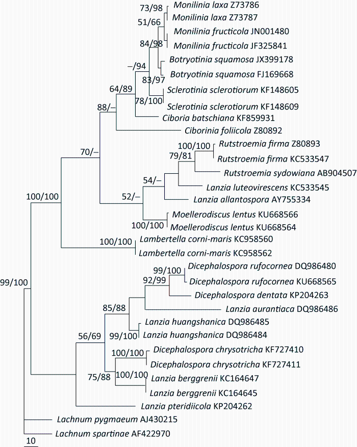
Based on the sequence analyses of ITS, Figure 1 showed the phylogenetic relationships among some fungi in Rutstroemiaceae and Sclorotiniaceae. The phylogeny suggested that the genus Lanzia seemed to be not monophyletic. Some of its species were associated with Moellerodiscus Henn. and Rutstroemia P. Karst., while others were related to Dicephalospora. Lanzia pteridiicola appeared as a separate lineage of the tree, and was not closely related to any species of Lanzia. Phylogenetic position of Lanzia might remain uncertain until sequences of its type species are available.
In this study, we concentrate on the Lanzia species closely related to Dicephalospora. Our results revealed that Lanzia aurantiaca and L. huangshanica were closely related to Dicephalospora rufocornea instead of other species of Lanzia (Fig. 1). These Lanzia species share many characteristics with Dicephalospora and its original generic concept except for the absence of a mucilaginous collar at each end of ascospores (Zhuang 1995), a feature thought to be critical or diagnostic for the genus when it was first established (Spooner 1987).
Fig. 1 Phylogenetic tree inferred from ITS sequences showing relationships among the selected rutstroemiaceous and sclerotiniaceous fungi. Statistical support values (≥50%) are shown at nodes (Left: MPBP, right: NJBP).
Our results indicate that two of the taxonomic criteria of Dicephalospora, the poles of ascospores capped with a mucilaginous collar and the J+ asci (Spooner 1987), do not carry phylogenetic information, and are not reliable features at generic level. To clarify some unsolved problems and to establish a clear concept of Dicephalospora, the genus needs to be emended to include those species possessing similar apothecial anatomic structure, producing substratal stromata, being closely related to Dicephalospora phylogenetically, but lacking of a mucilaginous collar at ends of ascospores. We transfer three Lanzia species to Dicephalospora.
Dicephalospora Spooner, Biblthca Mycol. 116: 267. 1987. emend. nov.
Stromata substratal, usually with blackened stromatized host tissues. Apothecia erumpent or superficial, stipitate, disc plane to convex, smooth, yellow, orange, dirty orange, orange-red, red or brown to blackish, receptacle concolorous with or lighter than the hymenium, stipe central, usually blackish at the base. Ectal excipulum of textura prismatica, hyphae with thickened, refractive walls, orientated at a very low angle to the receptacle surface. Medullary excipulum of textura intricata, hyphae hyaline, thin-walled. Subhymenium not clearly differentiated. Asci typically 8-spored, subcylindrical to cylindrical-clavate, J+ or J- in Melzer’s reagent. Ascospores hyaline, subelliptical, elliptical-fusoid to fusoid, smooth-walled, non-septate, guttulate, poles either capped with a mucilaginous collar or not. Paraphyses filiform, straight or somewhat curved at apex.
Dicephalospora aurantiaca (W.Y. Zhuang) W.Y. Zhuang & Z.Q. Zeng, comb. nov.
黄二头孢盘菌
Fungal Name FN 570254
≡Lanzia huangshanica f. aurantiaca W.Y. Zhuang, Mycosystema 7: 14. 1995.
≡Lanzia aurentiaca (W.Y. Zhuang) W.Y. Zhuang, in Zhuang & Liu, Mycotaxon 99: 127. 2007.
Specimens examined: China. Anhui, Huangshan, alt. 640m, 29-IX-1993, on stromatized leaf petioles of Castanopsis sp., Y.R. Lin, Y. Wang & W.Y. Zhuang 1149, HMAS 61847 (holotype); Anhui, Huangshan, alt. 800-1 300m, 26-IX-1993, on stromatized leaf petioles of Castanopsis sp., Y.R. Lin, Y. Wang & W.Y. Zhuang, S.M. Yu & W.J. Wu 1082, HMAS 61850 (paratype).
Notes: This fungus was previously treated as a member of Lanzia due to its absence of a mucilaginous collar at the poles of ascospores even though it is closely related to D. rufocornea (Zhuang 1995; Zhuang & Liu 2007). It should be a species of Dicephalospora since all the other features are congruent with one of the original species of the genus, which is also supported by the molecular data. The character, presence of mucilaginous collar at the poles of ascospores, should be treated as inter-specific variation instead of at generic level.
Dicephalospora calochroa (Syd.) Spooner, Biblthca Mycol. 116: 269. 1987.
二头孢盘菌
≡Orbilia calochroa Syd. & P. Syd., Bot. Jb. 54: 258. 1917.
We have not seen the specimen of this species. Wu et al. (1996) reported the fungus from Taiwan Province based on a collection from the Lianhua Pond in the catalogue of fungal specimens and cultures deposited in the National Museum of Natural Science (NMNS). The collection number is WAN 173, the herbarium number is NMNA F3436, the substrate and other collecting data were not mentioned.
Dicephalospora contracta W.Y. Zhuang, Xiao X. Liu & Z.Q. Zeng, sp. nov. Fig. 2
缩孢二头孢盘菌
Fungal Name FN 570253
Etymology: The specific epithet refers to the presence of a contraction in the middle of fusoid ascospores.
Apothecia convex to discoid, margin even, stipitate, 0.6-1mm in diam., with the stipe base dark; hymenium surface orange red when fresh, receptacle surface lighter. Ectal excipulum of textura prismatica, 30-64µm thick, cells 4-6×9-23µm, walls glassy or refractive. Medullary excipulum of textura intricata, 25-165µm thick, hyphae hyaline, thin-walled, somewhat refractive, 2-4µm wide. Subhymenium not distinguishable. Hymenium 120-130µm thick. Asci cylindrical-clavate, 8-spored, J+ in Melzer’s reagent, 99-114×7.5-8.5(-9)µm. Ascospores long-fusoid, constricted or narrower in the middle, more or less slipper-shaped, with a row of large guttules, hyaline, biseriate to irregularly biseriate, lacking of a gel cap at each end, 20-27×4-5µm. Paraphyses slightly enlarged and straight at apex, up to 3µm broad at apex and 1.5µm below, not or slightly exceeding the asci.
Fig. 2 Dicephalospora contracta (HMAS 271238). A-C: Dried apothecia on leaf petiole; D: Portion of hymenium showing paraphysis apices; E: Structure of apothecium near and at margin; F: Excipular structure; G-I: Ascospore; J-L: Sporoferous part of ascus showing ascospore arrangement. Scale bars: A=250µm, B=100µm, C=250µm, D=20µm, E=50µm, F=10µm, G-I=5µm, J-L=10µm.
Holotype: China, Yunnan, Pingbian, Daweishan, alt 1 900m, 4-XI-1999, on stromatized leaf petioles, W.Y. Zhuang & Z.H. Yu 3238, HMAS 271238.
Notes: Among the known species of the genus Dicephalospora, the new species is similar to D. rufocornea in apothecial color and the presence of a row of guttules in the fusoid ascospores, but D. rufocornea differs in larger apothecia with yellow to orange hymenium and lacking of a contraction in the middle of the longer ascospores (Spooner 1987; Zhuang 1998). Dicephalospora contracta is also similar to Lanzia huangshanica in ascospore size, but L. huangshanica differs obviously in the bright red hymenium surface, broader and shorter ectal excipular cells (8-22×5-9µm), broader asci (89-104×8-11µm), fusoid ascospores which are never constricted in the middle, and curved paraphysis apices.
It is very unfortunate that culture is not available for the new species. The type specimen is scanty, DNA was not extracted, and sequence data cannot be provided.
Dicephalospora damingshanica W.Y. Zhuang, Fungal Diversity 3: 190. 1999.
大明山二头孢盘菌
Specimen examined: China, Guangxi, Wuming, Damingshan, alt. 900m, 20-XII-1997, on rotten twig, W.P. Wu & W.Y. Zhuang 1860, HMAS 74893 (holotype).
Notes: The combined features, lemon-shape to elliptical-fusoid ascospores with a gel cap at each end and 22-32×9-12.7μm, make this fungus diagnostic in Dicephalospora (Zhuang 1999).
齿二头孢盘菌
Specimen examined: China, Yunnan, Mengla, alt. 650m, 17-X-1999, on rotten twig, Z.H. Yu & W.Y. Zhuang 3093, HMAS 266694 (holotype).
Notes: This fungus is the only known species of the genus having a dentate apothecial margin. It is closely related to D. rufocornea, especially similar to each other in the ascospore shape and size (Liu et al. 2015).
Dicephalospora huangshanica (W.Y. Zhuang) W.Y. Zhuang & Z.Q. Zeng, comb. nov.
黄山二头孢盘菌
Fungal Name FN 570252
≡Lanzia huangshanica W.Y. Zhuang & Korf, in Zhuang, Mycosystema 7: 13. 1995 [1994].
≡Lanzia huangshanica W.Y. Zhuang & Korf, in Zhuang, Mycosystema 7: 13. 1995 [1994] f. huangshanica.
Specimens examined: China. Anhui, Huangshan, alt. 640m, 29-IX-1993, on stromatized leaf petioles of Castanopsis sp., Y.R. Lin, Y. Wang & W.Y. Zhuang 1150, HMAS 61846 (holotype); Anhui, Huangshan, 11-VI-1994, on leaf petioles, Y.R. Lin, S.M. Yu, B. Deng, H.Y. Xing, W.B. Li & B.X. Liu HP 257, HMAS 68505 (paratype); Hunan, Yizhang, Mangshan, alt. 1 200m, 13-IV-2002, on stromatized leaf petioles, W.Y. Zhuang & Y.H. Zhang 4186, HMAS 97515.
Notes: This fungus is characterized by the red to dark red hymenium surface, curved paraphysis apices, and leaf petiole-inhabiting. It was also reported from Ilan County of Taiwan Province (Wang 1997) and with a beautiful color photograph of the fruitbody provided. Its distribution in China is probably wider than that we have known.
Dicephalospora phaeoparaphysis (W.Y. Zhuang) W.Y. Zhuang & Z.Q. Zeng, comb. nov.
暗丝二头孢盘菌
Fungal Name FN 570251
≡Lanzia phaeoparaphysis W.Y. Zhuang, Mycotaxon 56: 31. 1995.
Specimen examined: China. Jilin, Jiaohe, alt. 430m, 27-VIII-1991, on rotten leaf petioles, X.Q. Zhang & W.Y. Zhuang 735, HMAS 61867.
Notes: The apothecia of this fungus are about 0.7mm diam., and its hymenium surface is dirty orange to somewhat blackish when fresh due to the darkly pigmented contents inside the paraphyses. Its apothecial anatomy is very similar to that of D. huangshanica, as well as the shape and size of ascospores. However, D. huangshanica differs in the curved paraphyses apices with reddish content which makes the hymenium surface red to dark red.
Dicephalospora pinglongshanica W.Y. Zhuang, Fungal Diversity 3: 190. 1999.
平龙山二头孢盘菌
Specimen examined: China. Guangxi, Shangsi, Pinglongshan, alt. 500m, 2-I-1998, S.L. Chen, W.P. Wu & W.Y. Zhuang 2363, HMAS 74894 (holotype).
Notes: This fungus has small apothecia with hymenium surface yellow to reddish orange when fresh, J- asci, ascospores with a hyaline mucilaginous collar at both ends and 20-28×4.5-5.7μm, and straight paraphysis apices (Zhuang 1999). Among species of the genus, D. chrysotricha also possesses J- asci (Verkley 2004), whereas presence of characteristic hairs on receptacle surface obviously distinguishes this fungus from any other species of the genus.
Dicephalospora rufocornea (Berk. & Broome) Spooner, Biblthca Mycol. 116: 272. 1987.
橙红二头孢盘菌
≡Helotium rufocorneum Berk. & Broome, J. Linn. Soc., Bot. 14(74): 108. 1873.
≡Hymenoscyphus rufocorneus (Berk. & Broome) Dennis, Persoonia 3: 62. 1964.
≡Lanzia rufocornea (Berk. & Broome) Dumont, Mycotaxon 12: 272. 1980.
=Helotium subserotinum Henn. & E. Nyman, in Warburg, Monsunia 1: 33. 1899 [1900].
Specimens examined: China. Anhui, Huangshan, 11-IX-2007, on small twig, Y.R. Lin 1, HMAS 266585; Anhui, Huangshan, 12-IX-2007, on small twig, Y.R. Lin 4, HMAS 266586; Chongqing, Jinfoshan, 2-VI-2009, on rotten twig, B. Xiao 6108, 6109, Herbarium of Chongqing Institute of Medicinal Plant Cultivation; Guangdong, Dinghushan, alt. 50m, 9-X-1998, on twigs, W.Y. Zhuang & Z.H. Yu 2653, HMAS 82039; Guizhou, Daozhen, 19-VII-1988, on rotten wood, J.Z. Ying et al., HMAS 58264; Guizhou, Daozhen, 20-VII-1988, on rotten wood, Y. Li et al., HMAS 57797; Hainan, Baoting, 31-VII-1960, J.H. Yu & R. Liu, HMAS 29387; Hainan, Changjiang, Bawangling, alt. 1 100m, 6-XII-2000, on rotten wood, Y.H. Zhang, W.Y. Zhuang & Z.H. Yu 3666, HMAS 81346; Hainan, Changjiang, Bawangling, 07-XII-2000, on rotten wood, alt. 1 150m, W.Y. Zhuang, Z.H. Yu, & Y.H. Zhang 3668, HMAS 81347; Hainan, Ding’an, IX-1934, on twigs, X.K. Deng, HMAS 7204; Hainan, Ledong, Jianfengling, alt. 1 100m, 9-XII-2000, on twig, Y.H. Zhang, W.Y. Zhuang & Z.H. Yu 3748, HMAS 81345; Hainan, Ledong, Jianfengling, alt. 1 000m, 10-XII-2000, on rotten twig, Y.H. Zhang, W.Y. Zhuang & Z.H. Yu 3770, HMAS 81352; Hainan, Ledong, Jianfengling, alt. 1 000m, 10-XII-2000, on rotten twig, Z.H. Yu, W.Y. Zhuang & Y.H. Zhang 3795, HMAS 81353; Hainan, Lingshui, Diaoluoshan, 26-IX-1958, on rotten wood, R.Y. Zheng, HMAS 26780; Hainan, Linshui, Diaoluoshan, alt. 1 050m, 14-XII-2000, on twig, Z.H. Yu, Y.H. Zhang & W.Y. Zhuang 3841, HMAS 81348; Hainan, Linshui, Diaoluoshan, alt. 1 050m, 14-XII-2000, on petioles, Z.H. Yu, Y.H. Zhang & W.Y. Zhuang 3815, HMAS 81359; Hainan, Linshui, Diaoluoshan, alt. 1 050m, 14-XII-2000, on twig, Y.H. Zhang, W.Y. Zhuang & Z.H. Yu 3816, HMAS 81350; Hainan, Qiongzhong, Limushan, alt. 700m, 18-XII-2000, on twig, Y.H. Zhang, W.Y. Zhuang & Z.H. Yu, 3961, HMAS 81349; Hainan, Qiongzhong, Limushan, alt. 630m, 19-XII-2000, on twig, Y.H. Zhang, W.Y. Zhuang & Z.H. Yu, 4002, HMAS 81351; Hainan, Qiongzhong, Limushan, alt. 630m, 19-XII-2000, on petioles, Y.H. Zhang, W.Y. Zhuang & Z.H. Yu 4001, HMAS 81366; Hainan, Qiongzhong, Limushan, 18-XII-2000, on small twig, Y.H. Zhang, W.Y. Zhuang & Z.H. Yu, 3954, HMAS 81354; Hainan, Wuzhishan, alt. 850m, 16-XII-2000, on twig, X.M. Zhang et al. 3893, HMAS 81355; Hebei, Wulingshan, alt. 900m, 28-VIII-1989, on petiole of a tree leaf, B.C. Zhang [& W.Y. Zhuang] 535, HMAS 188430; Hong Kong, New territories, Tai Po Kao, 17-VII-1997, on wood, S.W. Wong, HKU(M) 7189; Hong Kong, New territories, Tai Po, 27-V-1998, C. Grgurinovic & J. Simpson, HKU(M) 10344; Hong Kong, New territories, Tai Po Kao, alt. 200m, 15-VII-1998, on rotten twigs, W.Y. Zhuang 2421, 2422, HKU(M) 10345, 10346, HMAS 74860, 74861; Hong Kong, New territories, Sai Kung, alt. 100m, 20-VII-1998, on rotten twig, W.Y. Zhuang 2442, HKU(M) 10358; Hong Kong, New Territories, Jubilee Reservoir, alt. 200m, 22-VII-1998, on rotten twigs and bark, W.Y. Zhuang 2458, HKU(M) 10361, HMAS 74862; Hong Kong, New territories, Tai Po Kao, alt. 200m, 22-VII-1998, on rotten twigs, W.Y. Zhuang 2468, HKU(M) 10364, HMAS 74863; Jiangxi, Jinggangshan, 23-X-1996, on twigs, W.Y. Zhuang & Z. Wang 1506, HMAS 71842; Jiangxi, Jinggangshan, alt. 1 300m, 24-X-1996, on twigs, W.Y. Zhuang & Z. Wang 1547, HMAS 71843; Shaanxi, Liuba, alt. 1 300m, 20-IX-1991, alt. 1 300m, on stromatized leaf petioles of Quercus sp., X.Q. Zhang & W.Y. Zhuang 827, HMAS 61843; Shaanxi, Foping, alt. 1 000m, 29-IX-1991, on stromatized leaf petioles of Quercus sp., W.Y. Zhuang 903, HMAS 61845; Shaanxi, Liuba, alt. 900m, 21-IX-1991, on stromatized leaf petioles of Quercus sp., W.Y. Zhuang 851, HMAS 61844; Sichuan, Qingchengshan, alt. 1 000m, 5-VII-1983, on twigs, W.Y. Zhuang 106, 109, HMAS 45081, 45082; Taiwan, Nantou, Lianhauchi, 17-05-1994, on bark, Y.Z. Wang, HMAS 61865; Taiwan, New Taipei City, 10-IX-2012, on small twigs, J.Y. Zhuang 10716, HMAS 266548; Yunnan, Kunming, 28-VIII-1938, S.Z. Zhao, HMAS 17064; Yunnan, Kunming, 09-VIII-1938, F.L. Tai, HMAS 17067; Yunnan, Kunming, [date unknown], [substrate unknown], C.C. Cheo, HMAS 17068; Yunnan, Kunming, 15-VII-1938, [substrate unknown], C.C. Cheo, HMAS 17069; Yunnan, Kunming, 16-VII-1938, [substrate unknown], C.C. Cheo, HMAS 17070; Yunnan, Kunming, 9-VII-1938, on withered twig, C.C. Cheo, HMAS 17073; Yunnan, Kunming, Xishan, 16-VII-1938, on withered twigs, C.C. Cheo, HMAS 17074; Yunnan, Kunming, 16-VII-1938, on withered twig, C.C. Cheo, HMAS 17075; Yunnan, Kunming, 27-VII-1938, [substrate unknown], C.C. Cheo, HMAS 17080; Yunnan, Kunming, 22-VII-1942, [substrate unknown], W.F. Chiu, HMAS 17081; Yunnan, Xishuangbanna, Menglun, alt. 500m, 16-IX-1987, J.Y. Zhuang, on twigs with bark, HMAS 68954; Yunnan, Xishuangbanna Botanical Garden, alt. 520m, 21-X-1988, on herbaceous stem, R.P. Korf, M. Zang, K.K. Chen & W.Y. Zhuang 201, HMAS 58762; Yunnan, Xishuangbanna, Limestone Hill, 22-X-1988, on herbaceous stems, R.P. Korf, M. Zang, K.K. Chen & W.Y. Zhuang 230, 232, 226, HMAS 68508, 68509, 72128; Yunnan, “51km” mark, road from Mengyang to Xishuangbanna Botanic Garden, alt. 600m, 23-X-1988, R.P. Korf, M. Zang, K.K. Chen & W.Y. Zhuang 273, HMAS 72129; Yunnan, Xishuangbann, “53km” mark, road from Mengyang to Xishuangbanna Botanical Garden, alt. 600m, 24-X-1988, on twigs, R.P. Korf, M. Zang, K.K. Chen & W.Y. Zhuang 304, HMAS 72175; Yunnan, Xishuangbanna, Endangered Plant Collections, alt. 520m, 25-X-1988, on twigs, R.P. Korf, M. Zang, K.K. Chen & W.Y. Zhuang 315, HMAS 72130.
Notes: This is a widely distributed fungus not only in China but also in the world. In China it has been reported from Anhui, Chongqing, Fujian, Guangxi, Guizhou, Hainan, Hebei, Hong Kong, Jiangsu, Jiangxi, Shaanxi, Sichuan, Taiwan and Yunnan (Teng 1963; Tai 1979; Korf & Zhuang 1985; Zhuang 1991, 1995, 1998; Wang 1997; Zhuang & Wang 1998a, 1998b).
1. Apothecial margin dentate ……………………...............................................…………...... D. dentata
1. Apothecial margin even ……………………………………..…………..…………..……………………………………. 2
2. Ascospores constricted in the middle ……………………..…………..………………………..... D. contracta
2. Ascospores not constricted in the middle ……..…………..………………………………………………………. 3
3. Ascospores 9-12.7μm wide ………………………..……………………...................... D. damingshanica
3. Ascospores less than 9μm wide…………………………………………………..………………….................... 4
4. Ascospores (27-)32-39×4-5.5(-6)μm …………………………....................……………. D. rufocornea
4. Ascospores shorter .............…………………………………………………..…………..…………….................. 5
5. Hymenium surface red or dark red …………………………………….................. D. huangshanica
5. Hymenium surface not red or dark red …………………………..…………..…………………….................. 6
6. Hymenium surface dirty orange to somewhat blackish when fresh ........ D. phaeoparaphysis
6. Hymenium surface of other colors ………………………..…………..………………………………………….... 7
7. Apothecia commonly less than 1mm diam. ..…………..…………………………………………………... 8
7. Apothecia larger ..…………..…………..…………………………………………………………………………………. 9
8. Apothecia about 1mm diam.; asci J+; paraphyses curved at apex …………… D. aurantiaca
8. Apothecia 0.3-0.6mm diam.; Asci J-; paraphyses straight at apex ….… D. pinglongshanica
9. Receptacle surface covered with hairs 20-50×3.5-5μm; asci J-; ascospores 14-21.5
(-27)×4-6.5μm..……..…………..……..………………………………………………………….…... D. chrysotricha
9. Receptacle surface without hairs; asci J+; ascospores 23-27(-29)×6.5-7.5μm … D. calochroa
Acknowledgements: The authors would like to thank Prof. Bo Xiao and other collector of the specimens for this study, the anonymous reviewer for valuable suggestions, and Ms. Xia Song for technique assistance.
The authors have declared that no competing interests exist.
| [1] |
Sclerotiniaceae XVI. On Helotium rufocorneum and Helotium fruternum. |
| [2] |
BioEdit: a user-friendly biological sequence alignment editor and analysis program for Windows 95/98/NT. |
| [3] |
Some new species and new records of discomycetes in China. |
| [4] |
|
| [5] |
TreeView: an application to display phylogenetic trees on personal computers. |
| [6] |
Helotiales of Australasia: Geoglossaceae, Orbiliaceae, Sclerotiniaceae, Hyaloscyphaceae. |
| [7] |
PAUP*: phylogenetic analysis using parsimony (*and other methods). version 4b10. Sinauer Associates ,
|
| [8] |
|
| [9] |
|
| [10] |
The ClustalX windows interface: flexible strategies for multiple sequences alignment aided by quality analysis tools. |
| [11] |
Redisposition of Chlorosplenium chrysotrichum to the genus Dicephalospora (Sclerotiniaceae, Ascomycota).
|
| [12] |
Designing primer sets for amplification of partial calmodulin genes from penicillia. |
| [13] |
Two species of the Sclerotiniaceae in Taiwan.
|
| [14] |
|
| [15] |
Catalogue of fungal specimens and cultures of NMNS. |
| [16] |
Some new species and new records of discomycetes in China. IV. |
| [17] |
A few petiole-inhabiting discomycetes in China.
|
| [18] |
|
| [19] |
Discomycetes of tropical China. VI. Additional species of Guangxi. |
| [20] |
Taxonomic reassessment of two taxa of helotialean cup-fungi. |
| [21] |
a. Discomycetes of tropical China. I. Collections from Hainan Island. |
| [22] |
b. Discomycetes of tropical China. II. Collections from Yunnan. |


/
| 〈 |
|
〉 |
