
庄文颖 , , 郑焕娣
ZHUANG Wen-Ying, ZHENG Huan-Di
通讯作者:
收稿日期: 2016-09-14
接受日期: 2016-12-29
网络出版日期: 2017-04-25
版权声明: 2017 中国科学院微生物研究所《菌物学报》编辑部 版权所有
展开
摘要
对中国科学院菌物标本馆保藏和近年来采集的我国小双孢盘菌属标本进行了分类研究,鉴定出14个种,包括7个新种(线孢小双孢盘菌、湖北小双孢盘菌、大孢小双孢盘菌、山地小双孢盘菌、蕨生小双孢盘菌、香地小双孢盘菌、中国小双孢盘菌)、4个中国新记录种(碘蓝小双孢盘菌、四孢小双孢盘菌、三隔小双孢盘菌、一个未定名种:Bisporella sp. 3999)和3个已报道种。对新种与相似种的主要形态学区别进行了详尽的比较和讨论,更新了部分种在我国的分布状况。提供了该属我国已知种的分种检索表。
关键词:
Abstract
Specimens on deposit in the Herbarium Mycologicum Academiae Sinicae and newly collected materials of the genus Bisporella from different regions of China were examined. Fourteen species are identified including seven new species (B. filiformis, B. hubeiensis, B. magnispora, B. montana, B. pteridicola, B. shangrilana, B. sinica), four new Chinese records (B. iodocyanescens, B. tetraspora, B. triseptata, Bisporella sp. 3999), and three previously recorded species. Comparisons between the new species and their allies are made. A key to the known species of the genus in China is provided.
Keywords:
The genus Bisporella Sacc. was established with Bisporella monilifera (Fuckel) Sacc. as the type species. The genus is characterized by apothecia discoid, cupulate to flat, stipitate or sessile, hymenium surface bright yellow, orange-yellow, pale yellow, whitish or beige; receptacle surface smooth, lighter than hymenium or concolorous; ectal excipulum of textura oblita, textura angularis or textura intricata, or a mixture of different tissues, tissue strongly gelatinous; medullary of textura intricata, hyphae hyaline; asci inoperculate, cylindrical-clavate, 4-8-spored, apical pores blue or not changing color in Melzer’s reagent; ascospores subfusoid, ellipsoidal-fusoid, elongate-ellipsoidal, fusoid with blunt ends or filiform, unicellular to multiseptate; and paraphyses filiform.
Korf & Carpenter (1974) pointed out that Bisporella is the correct name for the commonly used generic name Calycella Quél. and transferred four Calycella species to Bisporella. Regional studies of the genus were carried out, nomenclatural problems were solved, and new species have been described successively (Dennis 1956, 1978; Carpenter 1975, 1982; Beaton 1978; Korf 1982; Sharma & Korf 1982; Arendholz & Sharma 1983; Korf & Bujakiewicz 1985; Seifert & Carpenter 1987; Lizoň & Korf 1995; Gamundí & Romero 1998). According to Kirk et al. (2008), about 19 species are currently accepted.
Three Bisporella species were previously recorded in China (Teng 1963; Tai 1979; Korf & Zhuang 1985). In connection with our current work on the Chinese fungus flora, more materials are examined, and seven new species and four new Chinese records are reported. Our results indicate that China has a relatively high species diversity of Bisporella. Some species are common and widespread in the country, while others seem to be rare.
Because of the limitation of available sequence data, phylogenetic position of Bisporella in Helotiales and its relationships among other genera in Helotiaceae remains uncertain. In taxonomy of the genus, apothecial gross morphology, structure of ectal excipulum, and ascospore shape, size and septation are considered as important criteria for species identifications.
Specimens of Bisporella collected during 1988-2014 from different areas of China were examined, and deposited in the Herbarium Mycologicum Academiae Sinicae (HMAS). Dried apothecia were rehydrated in distilled water, and sectioned with a freezing microtome (YD-1508-III, Yidi Medical Appliance Factory, Jinhua, Zhejiang) at a thickness of ca. 15μm. Photographs were taken using a Canon G5 digital camera (Tokyo, Japan) connected to a Zeiss Axioskop 2 Plus microscope (Göttingen, Germany) for anatomical structures and a Leica M125 stereomicroscope (Wetzlar, Germany) for apothecial gross morphology. Terms used in descriptions followed Korf (1973). Measurements were made in sections or squash mounts using cotton blue as mounting medium. Cultures were not obtained when the specimens were collected, and sequences for very few species were available.
Genomic DNA was extracted from dried apothecia using the CTAB procedure (White et al. 1990) with minor modification. Sequences of rDNA ITS region were amplified and sequenced with the primer pair ITS1 and ITS4 (White et al. 1990). The PCR products were purified and sequenced at Beijing Tianyi Huiyuan Bioscience and Technology, China. Limited sequences of Bisporella from GenBank and newly obtained from a few Chinese collections were used to aid morphological considerations in the taxonomy.
Maximum parsimony (MP) and neighbor-joining (NJ) analyses were conducted via the program PAUP*4.0b10 (Swofford 2003) using parameters according to Zheng & Zhuang (2016). The topological confidence of NJ and MP trees was assessed by bootstrap analysis using 1 000 replications, each with 10 replicates of random stepwise addition of taxa.
线孢小双孢盘菌 图1
Bisporella filiformis W.Y. Zhuang & F. Ren, sp. nov. Fig. 1
Etymology: The specific epithet refers to the filiform ascospores.
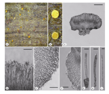
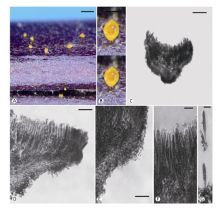
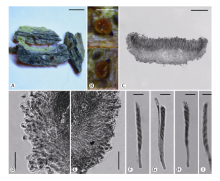
Fig. 1 Bisporella filiformis W.Y. Zhuang & F. Ren (HMAS 271290). A: Dried apothecia on natural substrate; B: Rehydrated apothecia; C: Median section of an apothecium; D: Portion of hymenium; E, F: Structure of ectal excipulum; G, H: Ascus; I: Ascospore. Scale bars: A=2mm; B=0.5mm; C=100μm; D=20μm; E-H=10μm; I=3μm.
Apothecia solitary to caespitose, discoid, sessile, about 0.5mm in diam.; hymenium surface yellow to light orange when dry, bright yellow when rehydrated, receptacle surface lighter than hymenium, smooth. Ectal excipulum of textura intricata to angularis, tissue gelatinous, 25-43μm thick, axes of cells near margin and at upper flank forming an acute angle and those at lower flank more or less perpendicular to receptacle surface, hyaline, 5-13×2-5μm. Medullary excipulum of textura intricata, 33-75μm thick, hyphae hyaline, 2-4μm wide. Subhymenium not distinguishable. Hymenium 60-75μm thick. Asci clavate, with apex slightly tapered, 8-spored, J+ in Melzer’s reagent with or without KOH pretreatment, 49-61×5.5-7.5μm. Ascospores filiform with both ends somewhat narrow or vermiform, hyaline, smooth, multi-septate (6-10), guttulate, 30-42×1.8-2μm, arranged in a fascicle in ascus. Paraphyses filiform, septate, often branched at apical portion, 1.5-2.5μm wide at apical portion and 1.2-1.5μm below.
Holotype: China. Hainan, Ledong, Jianfengling, alt. 1 100m, 9-XII-2000, on twig, W.Y. Zhuang & Z.H. Yu 3732, HMAS 271290.
Notes: The new species shares the common features of Bisporella, such as apothecia yellow, tissue gelatinous, axes of ectal excipular cells forming an acute angle to receptacle surface, and on woody substrates. Among species of the genus (Dennis 1956, 1958; Korf & Carpenter 1974; Carpenter 1975; Beaton 1978; Sharma & Korf 1982; Seifert & Carpenter 1987; Galán 1988; Gamundí & Romero 1998), the new species is diagnostic by its filiform to vermiform ascospores with 6-10 septa, which has not been reported in any known species of the genus.
It is unfortunate that strain was not obtained when the fungus was collected, and DNA sequences are not available.
湖北小双孢盘菌 图2
Bisporella hubeiensis H.D. Zheng & W.Y. Zhuang, sp. nov. Fig. 2
Etymology: The specific epithet refers to the type locality.
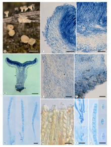
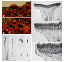
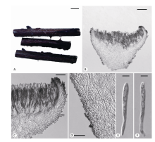
Apothecia two or three in a cluster or solitary, discoid, drying concave, stipitate, 0.3-0.9mm in diam.; hymenium surface pure white, drying pale yellow; receptacle surface drying yellowish white, slightly villiform; stipe cylindrical, whitish to concolorous with receptacle. Ectal excipulum of textura prismatica to intricata, hyphae oriented at a moderate angle to receptacle surface, tissue gelatinous, 38-82μm thick, cells hyaline, 10-20×2.5-5μm, the ends of terminal cells slightly swollen. Medullary excipulum of textura intricata, 30-70μm thick, hyphae hyaline, 2-3μm wide. Subhymenium not distinguishable or poorly developed. Hymenium 70-80μm thick. Asci arising from croziers, cylindrical-clavate, with apex rounded, 8-spored, J+ in Melzer’s reagent without KOH pre-treatment, 50-60×3.5-4.4μm. Ascospores subellipsoidal, hyaline, smooth, non-septate, with two large guttules, 4.6-6×2.2-2.8μm, uniseriate. Paraphyses cylindrical, with apex rounded, hyaline, septate, unbranched, 1.5-2μm wide.
Holotype: China. Hubei, Shennongjia, Jinhouling, alt. 1 800m, 16-IX-2004, on decorticated wood, W.Y. Zhuang 5735, HMAS 271402.
Notes: Among species of the genus, this fungus is similar to B. subpallida (Rehm) Dennis in color of apothecia and size of asci (Dennis 1956). However, the latter fungus has smooth receptacle surface instead of slightly villiform, a different ectal excipular structure that the hyphae are parallel-arranged, and longer ascospores (6-10×2.5-3μm) which are biseriately arranged in the ascus.
It is unfortunate that strain was not obtained when the fungus was collected, and DNA sequences are not available.
Fig. 2 Bisporella hubeiensis H.D. Zheng & W.Y. Zhuang (HMAS 271402). A: Dried apothecia on natural substrate; B: Longitudinal section of apothecium; C: Excipular structure at margin; D: Excipular structure at flank; E: Structure in the middle of stipe; F: Structure of stipe base; G: Asci; H: Ascus apical rings in iodine; I: Croziers at ascus base; J: Ascospores. Scale bars: A=0.5mm; B=200μm; C-F=20μm; G=10μm; H-J=5μm.
大孢小双孢盘菌 图3
Bisporella magnispora W.Y. Zhuang & H.D. Zheng, sp. nov. Fig. 3
Etymology: The specific epithet refers to the large ascospores.
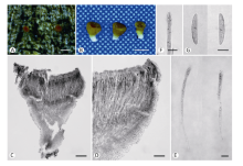
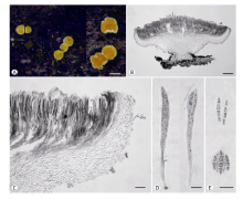
Apothecia solitary, discoid, with margin even, stipitate, 0.4-0.9mm in diam.; hymenium white when fresh, receptacle concolorous, surface smooth. Ectal excipulum of textura oblita, 20-50μm thick, tissue highly gelatinous, hyphae hyaline, parallel-arranged and forming an acute angle to the outer surface, 4-6.5μm wide. Medullary excipulum of textura intricata, 25-130μm thick, hyphae hyaline ca. 2μm thick. Subhymenium not distinguishable. Hymenium 240-250μm thick. Asci arising from croziers, elongate-clavate or narrowly clavate, 4-8-spored, J+ in Melzer’s reagent and pore walls strongly blue, 229-254×9-11μm. Ascospores elongate-fusoid with blunt ends, hyaline, unicellular, with a dark-stained area in cotton blue, 26-37.5×4.5-6μm, irregularly biseriate. Paraphyses filiform, 2-2.5μm wide.
Holotype: China. Hubei, Wufeng, Houhe Nature Reserve, alt. 800m, 13-IX-2004, on rotten wood, W.Y. Zhuang & C.Y. Liu 5601, HMAS 275575.
Notes: The apothecial anatomy of this fungus is somewhat similar to that of Bisporella tetraspora (Feltgen) S.E. Carp., but its asci are very long and contain usually 4-8 ascospores instead of 4 spores at maturity, and ascospores are very larger, 26-37.5×4.5-6μm instead of 9-13×3.5-4μm in B. tetraspora. It represents obviously a huge-spored new species.
Fig. 3 Bisporella magnispora W.Y. Zhuang & H.D. Zheng (HMAS 275575). A: Dried apothecia on natural substrate; B: Dried apothecia; C: Structure of apothecium; D: Excipular structure at margin and flank; E: Asci; F: Portion of an ascus showing arrangement of ascospores; G: Ascospores. Scale bars: A, B=0.5mm; C=100μm; D=50μm; E, F=20μm; G=10μm.
It is unfortunate that strain was not obtained when the fungus was collected, and DNA sequences are not available.
山地小双孢盘菌 图4
Bisporella montana W.Y. Zhuang & H.D. Zheng, sp. nov. Fig. 4
Etymology: The specific epithet refers to the occurrence in mountain areas.
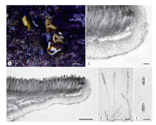
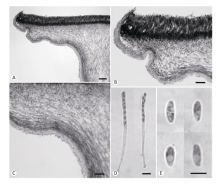
Apothecia discoid, stipitate, 1-3mm in diam.; hymenium surface yellow, receptacle surface paler, smooth. Ectal excipulum of textura intricata, sometimes mixed with textura oblita especially at margin, tissue highly gelatinized, 38-84μm thick, hyphae hyaline, refractive, 2.5-4.5μm wide. Medullary of textura intricata, tissue not gelatinized, 38-460μm thick, hyphae hyaline, 1.5-2.5μm wide. Subhymenium not distinguishable or very poorly developed, 0-13μm thick. Hymenium 100-115μm thick. Asci arising from crosiers, subcylindrical above and tapering towards the base, 8-spored, J- in Melzer’s reagent or iodine reaction very difficult to detect, 88-114×4.5- 5.5μm.Ascospores ellipsoidal to ellipsoidal with narrow ends, hyaline, smooth, non- to uni-septate, with two small polar guttules, 5.5-6.5(-8.5)×2.5- 3(-3.3)μm, uniseriate. Paraphyses filiform, ca. 1.5μm wide.
Fig. 4 Bisporella montana W.Y. Zhuang & H.D. Zheng (HMAS 275566). A: Dried apothecia on natural substrate; B: Structure of apothecium; C: Excipular structure at margin and flank; D: Asci; E: Ascospores. Scale bars: A=0.5mm; B=20μm; C=100μm; D=10μm; E=5μm.
Holotype: China. Yunnan, Pingbian, Daweishan, alt. 1 900m, 5-IX-1999, on rotten wood, W.Y. Zhuang & Z.H. Yu 3325, HMAS 275566.
Other specimen examined: China. Yunnan Pingbian Daweishan, alt. 1 900m, 5-IX-1999, on hard wood, W.Y. Zhuang & Z.H. Yu 3319, HMAS 275567.
Sequence data: ITS rDNA (HMAS 275566) - GenBank acc. no. KX781358.
Notes: This species is similar to Bisporella citrina (Batsch) Korf & S.E. Carp. in excipular structure. Detailed comparisons reveal that B. citrina has larger apothecia (0.7-7mm diam.), asci [90-155×5-7.5(-8.5)μm] and ascospores [9-13.5(-14)×3-4(-4.5)μm]. The ascospore shapes
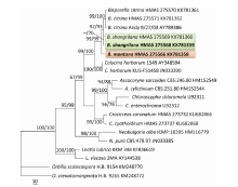
are ellipsoidal-fusoid and with two large guttules in B. citrine, and ellipsoidal or ellipsoidal with narrow ends and containing two small polar guttules in B. montana. In addition, 18-21bp sequence differences are found in rDNA ITS regions between the two fungi. Sequence analyses support the separation of them at species level (Fig. 8).
蕨生小双孢盘菌 图5
Bisporella pteridicola F. Ren & W.Y. Zhuang, sp. nov. Fig. 5
Fig. 5 Bisporella pteridicola F. Ren & W.Y. Zhuang (HMAS 271269). A: Dried apothecia on natural substrate; B: Rehydrated apothecia; C: Structure of apothecium; D, E: Excipular structure; F: Portion of hymenium; G: Asci. Scale bars: A=1mm; B=0.5mm; C=50μm; D, E=20μm; F, G=10μm.
Etymology: The specific epithet refers to the substrate.
Apothecia discoid to flat, sessile, 0.1-0.9mm in diam.; hymenium surface beige, beige-yellow to yellow when fresh and bright yellow when dry, receptacle surface concolorous or slightly lighter, nearly smooth. Ectal excipulum of textura angularis, 18-46μm thick, tissue gelatinized, axis of cells with a low angle to outer surface, some of the outer most cells slightly protruding from outer surface, cells hyaline, 2-6μm wide. Medullary of textura intricata, tissue not gelatinous, 27-45μm thick, hyphae hyaline, 2.5-4μm wide. Subhymenium 9-12μm thick. Hymenium 45-55μm thick. Asci arising from crosiers, clavate, tapering towards the base, 8-spored, J- in Melzer’s reagent, 36-46×5-6.5μm. Ascospores subellipsoidal to subfusoid, hyaline, smooth, unicellular, with two large guttules, 7-9×2-3.5μm, biseriate. Paraphyses filiform, 1.2-1.5μm wide.
Holotype: China. Guangdong, Zhaoqing, Dinghushan, alt. 150m, 10-X-1998, on fern rachis, W.Y. Zhuang & S.L. Chen 2682, HMAS 271269.
Other specimen examined: China. Guangdong, Fengkai, Heishiding, alt. 300m, 27-X-1998, on fern rachis, W.Y. Zhuang & Z.H. Yu 2852, HMAS 271270.
Notes: Bisporella pteridicola is the only species of the genus growing on fern rachis other than seed plants. Its apothecial structure fits well the generic concept of Bisporella. The cells in the ectal excipulum are angular, which is similar to that of B. iodocyanescens Korf & Bujak. But asci of the latter fungus are larger (57-71×4.5-6.5μm), ascospores are shorter (4.8-7×2-3.5μm), subhymenium turns blue in Melzer’s reagent, and not occurring on fern.
It is unfortunate that strain was not obtained when the fungus was collected and DNA sequences are not available.
香地小双孢盘菌 图6
Bisporella shangrilana W.Y. Zhuang & H.D. Zheng, sp. nov. Fig. 6
Etymology: The specific epithet refers to the type locality.
Apothecia discoid, stipitate, 1-3mm in diam.; hymenium surface orange-yellow, receptacle surface paler, smooth. Ectal excipulum of textura oblita at margin and textura intricata at flank, tissue gelatinous, 15-25μm thick, hyphae hyaline, 2-4μm wide. Medullary of textura intricata, tissue not gelatinous, 50-560μm thick or thicker, hyphae hyaline, 1.5-3μm wide. Subhymenium not distinguishable. Hymenium 110-125μm thick. Asci arising from crosiers, subcylindrical above and tapering towards the base, 8-spored, J+ in Melzer’s reagent as two blue lines, 88-105×4.5-5.5μm. Ascospores ellipsoidal to ellipsoidal with narrow ends, hyaline, smooth, non- to uni-septate, with two small polar guttules, 5-7.2(-8)×(2.2-)2.5-3.3μm, uniseriate. Paraphyses filiform, ca. 1.5μm wide.
Holotype: Yunnan, Shangrila, Bitahai, alt. 3 800m, 12-VIII-2008, on rotten wood, X.Q. Zhang & D.Z. Ren 7345, HMAS 275568.
Other specimen examined: Yunnan, Shangrila, Bitahai, alt. 3 800m, 12-VIII-2008, on dead twig, X.Q. Zhang & D.Z. Ren 7322, HMAS 275569.
Sequence data: ITS rDNA (HMAS 275568, 275569) - GenBank acc. nos. KX781359, KX781360.
Notes: Bisporella shangrilana is very similar to B. montana in sizes of apothecia, asci and ascospores. However, they are completely different in excipular structure which is highly gelatinous and
much thicker (38-84μm thick) in B. montana but less refractive and extremely thin in B. shangrilana. The ascus apex iodine reaction is also different, which is not blue in B. montana but dark blue in B. shangrilana. As sequence data are compared, ITS sequences of the two collections of B. shangrilana are identical, while 11bp are different in rDNA ITS region between the two fungi. The sequence analyses of ITS support the recognition of B. shangrilana as a new species (Fig. 8).
Fig. 6 Bisporella shangrilana W.Y. Zhuang & H.D. Zheng (HMAS 275568). A, B: Dried apothecia on natural substrate; C: Structure of apothecium; D, E: Excipular structure at margin and flank; F: Asci; G: Ascospores. Scale bars: A, B=0.5mm; C=100μm; D, E=50μm; F=20μm; G=10μm.
中国小双孢盘菌 图7
Bisporella sinica W.Y. Zhuang, sp. nov. Fig. 7
Etymology: The specific epithet refers to the occurrence of the fungus in China.
Apothecia gregarious, sometimes confluent, discoid, sometimes with center convex, stipitate, 1.5-5mm in diam.; hymenium surface bright yellow to orange-yellow when fresh, receptacle surface concolorous or slightly lighter, smooth. Ectal excipulum
Fig. 7 Bisporella sinica W.Y. Zhuang (HMAS 271337). A: Structure of apothecium; B: Structure of apothecium at margin and flank; C: Structure of apothecium near stipe; D: Asci; E: Ascospores. Scale bars: A=100μm; B, C=50μm; D=10μm; E=5μm.
of textura prismatica to angularis at margin and upper flanks, of textura intricata at lower flank, tissue gelatinous, 23-38(-45)μm thick, cells and hyphae hyaline, cells 3-15×4-6μm, hyphae 2-4μm wide. Medullary excipulum of textura intricata, tissue not gelatinous, 38-360μm thick, hyphae hyaline, 1.5-3(-4)μm wide. Subhymenium not well-developed. Hymenium 94-115μm thick. Asci arising from croziers, clavate, tapering towards the base, 8-spored, J+ in Melzer’s reagent, 82-110(-115)×5-6.5(-7)μm. Ascospores elongate- ellipsoidal, hyaline, smooth, uni-septate at maturity, guttulate, 6-9.5(-10)×3-4μm, uniseriate. Paraphyses filiform, septate, 1.5-2μm wide.
Holotype: China. Jilin, Jiaohe, alt. 450m, 27-VIII-1991, on rotten wood, F.Y. Bai & W.Y. Zhuang 740, HMAS 271337.
Other specimens examined: China. Gansu, Tewo, Baiyun Forestry Station, alt. 2 300m, 12-X-1992, on rotten wood, W.Y. Zhuang 1028, HMAS 271342; Jilin Jiaohe, alt. 450m, 29-VIII-1991, on rotten wood, W.Y. Zhuang & J.B. Chen743, HMAS 271338; Jilin, Jiaohe, alt. 550m, 31-VIII-1991, on wet rotten wood, W.Y. Zhuang 793, HMAS 271339; Jilin, Jiaohe, Shimenling, 29-VIII-1991, on rotten wood, W.Y. Zhuang 775, HMAS 271340; Shaanxi, Foping, alt. 1 300m, 26-IX-1991, on rotten wood, W.Y. Zhuang 882, HMAS 271341; Sichuan, Daofu, alt. 3 780m, 27-VIII-1997, on rotten wood, Z. Wang 2171, HMAS 72810; Sichuan, Daofu, alt. 3 600m, 30-VIII-1997, on rotten wood, Z. Wang 2192, HMAS 74617; Sichuan, Gonggashan, alt. 1 900m, 14-VIII-1997, on rotten wood, Z. Wang 2009, HMAS 72028; Sichuan, Gonggashan, alt. 1 900m, 16-VIII-1997, on rotten wood, Z. Wang 2013, HMAS 72049; Sichuan, Xiangcheng, Dongwang, alt. 3 800m, 26-VII-1997, on rotten wood, Z. Wang 200, HMAS 76077; Xinjiang, Hemuxiang, alt. 1 100m, 6-VIII-2003, on rotten wood, W.Y. Zhuang & Y. Nong 4750, HMAS 275572.
Notes: This species is relatively common in China. It is characterized by apothecia often confluent, ectal excipulum at margin and upper flank of textura prismatica, and axis of ectal excipular cells with an acute angle to the outer surface. It is similar to Bisporella confluens (Sacc.) Korf & Bujak. from North America in apothecial size, cell arrangement of ectal excipulum at margin and upper flank. However, they are different in the anatomic structure at lower flank and sizes of asci and ascospores. Bisporella confluens has asci 125-135×7.5-8.8μm, ascospores 11.3-14.2(-16.5)×3.3-4.4(-4.7)μm, and larger apothecia which are occasionally up to 3cm in diameter (Korf & Bujakiewicz 1985).
The fungus is somewhat similar to B. oritis Beaton from Australia in excipular structure at and near the apothecial margin. However, B. oritis has smaller apothecia ca. 1mm in diam., smaller asci 60-75×6.5-7.5μm and J- in iodine, and smaller ascospores 7-9.5×3.5-4μm (Beaton 1978). They are not conspecific.
Fig. 8 Maximum parsimony tree inferred from ITS sequences showing taxonomic positions of Bisporella montana and B. shangrilana. Bootstrap values (≥50%) of maximum parsimony (left) and neighbor-joining (right) are indicated at the nodes.
The new species slightly resembles B. schusteri Gamundí from Argentina in size of apothecia (1-4mm in diam.) and length of asci (95-115×3.6-5.4μm). But B. schusteri has J- asci which are narrower, larger ascospores [(8-)9- 11(-14.5)×3.6-4.5μm], and a quite different ectal excipular structure (Gamundí & Romero 1998). They are distinguishable at species level.
Some of the Chinese collections were previously filed under B. citrina or B. sulfurina (Quél.) S.E. Carp.
碘蓝小双孢盘菌 图9
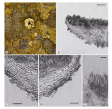
Bisporella iodocyanescens Korf & Bujak., Agarica 6(12): 304, 1985. Fig. 9
Apothecia discoid to flat, sessile, ca. 1mm in diam.; hymenium surface yellow to orange-yellow when fresh, receptacle surface concolorous, nearly smooth. Ectal excipulum of textura angularis to globulosa, tissue gelatinous, 27-41μm thick, cells hyaline, walls slightly thickened and refractive, 4-11μm in diam. Medullary excipulum of textura intricata, 27-75μm thick, hyphae hyaline, 3-4.5μm wide. Subhymenium turning light blue in Melzer’s reagent, 11-15μm thick. Hymenium 60-80μm thick. Asci arising from croziers, cylindric-clavate, tapering towards the base, 8-spored, J+ in Melzer’s reagent with KOH pretreatment, 57-71×4.5-6.5μm. Ascospores subellipsoidal, hyaline, smooth, unicellular, 4.8-7×2-3.5μm, uniseriate. Paraphyses filiform, 1-2μm wide.
Fig. 9 Bisporella iodocyanescens Korf & Bujak. (HMAS 271265). A: Dried apothecia on natural substrate; B: Rehydrated apothecia; C: Structure of apothecium; D, E: Structure of excipulum; F-I: Asci. Scale bars: A=4mm; B=1mm; C=100μm; D, E=20μm; F-I=10μm.
Specimen examined: China. Xinjiang, Xinyuan, Nalati, alt. 2 200m, 15-VIII-2003, on rotten wood, W.Y. Zhuang & Y. Nong 4959, HMAS 271265.
Notes: This is a rare fungus. The type locality is New York, USA. A single collection was found from China. Compared with the American material, the Chinese collection has slightly larger asci (57-71×4.5-6.5μm vs. 55-66×3.6-4μm) and ascospores (4.8-7×2-3.5μm vs. 4.5-6.3×1.5-2μm) (Korf & Bujakiewicz 1985) which are treated as infraspecific variations. The most diagnostic features of the species are the ectal excipulum of textura angularis to textura globulosa and medullary excipulum turning blue in Melzer’s reagent.
四孢小双孢盘菌 图10
Bisporella tetraspora (Feltgen) S.E. Carp., Mem. New York Bot. Gdn. 33: 262, 1981. Fig. 10
Phialea tetraspora Feltgen, Vorstud Pilzfl. Luxemb., Nachtr. 2: 51, 1901.
Fig. 10 Bisporella tetraspora (Feltgen) S.E. Carp. (HMAS 271285). A: Dried apothecia on natural substrate; B: Structure of apothecium; C: Structure of apothecium at margin and flank; D: Ectal excipulum at lower flank; E, F: Ascus. Scale bars: A=5mm; B=100μm; C=30μm; D=20μm; E, F=10μm.
Apothecia discoid to flat, sessile to substipitate, 0.2-0.8mm in diam.; hymenium surface dirty white to yellow, receptacle surface concolorous or slightly lighter, nearly smooth. Ectal excipulum of textura oblita to intricata, tissue gelatinous, 25-50μm thick, hyphae hyaline, 2-4μm wide. Medullary of textura intricata, 20-115μm thick, hyphae hyaline, 2.5-4μm wide. Subhymenium not distinguishable. Hymenium 70-80μm thick. Asci arising from croziers, cylindric-clavate, tapering towards the base, 4-spored, J+ in Melzer’s reagent, 60-78×5.5-8.5μm. Ascospores fusiform-ellipsoid, hyaline, smooth, unicellular, with 2 large guttules, 9-13×3.5-4.5μm, uniseriate. Paraphyses filiform, septate, 1.2-2μm wide.
Specimens examined: China. Gansu, Tewo, Luoda Forestry Farm, alt. 2 100m, 31-VII-1998, on dead twig, S.L. Chen 125b, HMAS 275573; Sichuan, Barkam, alt. 2 939m, 30-VII-2013, on withered twig, Z.Q. Zeng, Z.X. Zhu & F. Ren 8482, 8487, HMAS 271285, 271286.
Notes: This species is different from any other species of the genus by its 4-spored asci. It has been reported from Turkey, Canada, Mexico, United States and Columbia.
三隔小双孢盘菌 图11
Bisporella triseptata (Dennis) S.E. Carp. & Dumont, Caldasia 12(no. 58): 344, 1978. Fig. 11
Calycella sulfurina var. triseptata Dennis, Kew Bull. 14: 431, 1960.
Fig. 11 Bisporella triseptata (Dennis) S.E. Carp. & Dumont (HMAS 275574). A: Dried apothecia on natural substrate; B: Structure of apothecium; C: Excipular structure at margin and flank; D: Asci; E: Ascospores. Scale bars: A=0.5mm; B=100μm; C=20μm; D, E=10μm.
Apothecia discoid, margin even, substipitate, 0.3-0.8mm in diam., hymenium light yellow, receptacle concolorous. Ectal excipulum of textura oblita to intricata, tissue highly gelatinous, 40-150μm thick, hyphae nearly parallel-arranged, 3-5μm wide. Medullary excipulum of textura intricata, with two layers, the outer layer gelatinous, not easily separated from ectal excipulum, 50-60μm thick, the inner layer not gelatinous, 35-100μm thick, hyphae 2-3μm wide. Asci clavate, with a simple base, 8-spored, J- in Melzer’s reagent, 78-90×6.6-8μm. Ascospores fusoid, mostly 3-septate, hyaline, 12-17×2.5-3.5μm, irregularly biseriate. Paraphyses filiform, 2-2.5μm wide.
Specimen examined: China. Gansu, Tewo, Lazikou Forestry Farm, 2 200-2 800m, 26-VII-1998, on small culm of bamboo, S.L. Chen 98a, HMAS 275574.
Notes: The species was previously reported from South America. The Chinese collection extends its distribution to Asia. The Chinese material possesses somewhat wider asci and smaller apothecia, which are treated as infraspecific variations (Carpenter & Dumont 1987).
小双孢盘菌属未定名种3999 图12
Bisporella sp. 3999 Fig. 12
Fig. 12 Bisporella sp. 3999 (HMAS 271268). A: Dried apothecia on natural substrate; B: Structure of apothecium; C, D: Excipular structure; E: Portion of hymenium. Scale bars: A=2mm; B=50μm; C, D=10μm; E=20μm
Apothecia solitary to gregarious, discoid to flat, sessile, 0.5-0.8mm in diam.; hymenium surface dirty white to yellow when fresh, receptacle surface slightly lighter than hymenium, nearly smooth. Ectal excipulum of textura oblita, 27-47μm thick, tissue gelatinous, cells hyaline, 6-11×1.5-3.7μm. Medullary excipulum of textura intricata, 25-60μm thick, hyphae hyaline, 2-4μm wide. Subhymenium not distinguishable or poorly developed. Hymenium 39-47μm thick. Asci cylindrical-clavate, tapering towards the base, 8-spored, J+ in Melzer’s reagent and pore walls turning light blue, 30-35×4.5-5μm. Ascospores ellipsoidal-fusoid, hyaline, smooth, unicellular, 4.8-5.8×1.5-2.5μm, irregularly uniseriate. Paraphyses filiform, 1.2-1.8μm wide.
Specimen examined: China. Hainan, Qiongzhong, Limushan, alt. 700m, 15-XII-2000, on bark, W.Y. Zhuang, Y.H. Zhang, Z.H. Yu 3999, HMAS 271268.
Notes: Compared with the known species of the genus, HMAS 271268 possesses very small asci and ascospores. It represents a new species but the material is too scanty to be a type. We tentatively treat it as “Bisporella sp. 3999”.
橘色小双孢盘菌
Bisporella citrina (Batsch) Korf & S.E. Carp., Mycotaxon 1: 58, 1974.
Peziza citrina Batsch, Elench. Fung., cont. sec. (Halle): 95, 1789.
= Octospora citrina Hedw., Descr. Micr.-anal. Musc. Frond. 2: 28, tab. 8B, figs. 1-7, 1789.
Helotium citrinum (Hedw.) Fr., Summa Veg. Scand., Section Post. (Stockholm): 355, 1849. Teng, Fungi of China p. 266, 1963.
Calycella citrina (Hedw.) Boud., Bull. Soc. Mycol. Fr. 1: 112, 1885. Tai, Sylloge Fungorum Sinicorum p. 92, 1979.
Specimens examined: China. Gansu, Tewo, alt. 2 600m, 10-IX-1992, on rotten wood, X.Q. Zhang & W.Y. Zhuang 1010, HMAS 271324; Gansu, Zhouqu, Shatan Forestry Station, alt. 2 430m, 3-IX-1992, on wet rotten wood, X.L. Mao & W.Y. Zhuang 942, HMAS 275556; Heilongjiang, Dailing, Liangshui Forestry Station, alt. 400-500m, 28-VIII-1996, on rotten wood, W.Y. Zhuang & Z. Wang 1291, HMAS 271314; Heilongjiang, Dailing, Liangshui Forestry Station, alt. 400m, 31-VIII-1996, on rotten wood, W.Y. Zhuang & Z. Wang 1369, 1370, 1372, 1378, 1379, HMAS 271315, 271316, 271317, 271318, 271319; Hubei, Shennongjia, Huangbaoping, alt. 1 750m, 16-IX-2014, on rotten wood, H.D. Zheng, Z.Q. Zeng, W.T. Qin, K. Chen 9636, HMAS 275571; Hubei, Shennongjia, Jinhouling, alt. 2 500m, 14-IX-2014, on rotten wood, H.D. Zheng, Z.Q. Zeng, W.T. Qin, K. Chen 9516, HMAS 275578; Hubei, Wufeng, Houhe Nature Reserve, alt. 800m, 12-IX-2004, on rotten wood, W.Y. Zhuang & C.Y. Liu 5510, HMAS 271320; Jilin, Dunhua, alt. 800m, 15-VIII-2000, on rotten twig, W.Y. Zhuang, Z.H. Yu & Y.H. Zhang 3491, HMAS 271309; Jilin Jiaohe, Dadingzishan, alt. 800m, 26-VIII-1991, on rotten wood, W.Y. Zhuang 764, HMAS 271310; Jilin Jiaohe, Jiaohe Forestry Station, Shimenling, 29-VIII-1991, on wet rotten wood, W.Y. Zhuang 772, HMAS 271311; Jilin Jiaohe, Erdaohe, alt. 700m, 1-IX-1991, on wet rotten wood, W.Y. Zhuang 809, 810, HMAS 271312, 271313; Qinghai, Huzhu, Beishan, alt. 2 800-3 000m, 15-VIII-2004, on rotten wood, W.Y. Zhuang & C.Y. Liu 5330, HMAS 271325; Shaanxi, Foping, Tianhuashan, alt. 1 300m, 23-IX-1991, on wet rotten wood, W.Y. Zhuang 888, HMAS 271322; Shaanxi, Foping, alt. 1 300m, 26-IX-1991, on wet rotten wood, W.Y. Zhuang 892, HMAS 271323; Sichuan, Gonggashan, alt. 1 900m, 14-VIII-1997, on rotten bark, D. Hibbet & Z. Wang 2006, HMAS 72026; Sichuan, Gonggashan, alt. 1 900m,1997 VIII 14,on rotten wood, Z. Wang 2014, HMAS 72029; Sichuan, Gonggashan, alt. 3 300m,8-IX-1997, on rotten wood, Z. Wang 2237, HMAS 74611; Sichuan, Gonggashan, Hailuogou, alt. 1 700-1 900m, 17-VIII-1997, Z. Wang 2059, HMAS 72040; Sichuan, Jiuzhaigou, 18-IX-1992, on rotten wood, W.Y. Zhuang 1048, HMAS 271321; Sichuan, Xiangcheng, alt. 4 200m, 24-VII-1998, on rotten wood, Z. Wang 155, HMAS 76098; Xinjiang, Altaishan, alt. 1 100m, 7-VIII-2003, on rotten wood, W.Y. Zhuang & Y. Nong 4798, 4799, HMAS 271326, 271327; Xinjiang, Altaishan, alt. 1 250m, 9-VIII-2003, on rotten twig, W.Y. Zhuang & Y. Nong 4823, 4824, HMAS 271328, 271329; Xinjiang, Bourgin, Hemuxiang, alt. 1 100m, 5-VIII-2003 VIII 5, on rotten wood, W.Y. Zhuang & Y. Nong 4686, 4691, 4699, HMAS 271330, 271331, 271332; Xinjiang, Bourgin, Hemuxiang, alt. 1 100m, 6-VIII-2003, on bark, W.Y. Zhuang & Y. Nong 4751, HMAS 271333; Xinjiang, Kanas, alt. 1 300m, on rotten wood, 8-VIII-2003, W.Y. Zhuang & Y. Nong 4806, HMAS 271334; Yunnan, Hekou, Daweishan, alt. 1 900m, 5-XI-1999, on rotten bark, W.Y. Zhuang & Z.H. Yu 3300, HMAS 275579; Yunnan, Pingbian, Daweishan, alt. 1 900m, 4-XI-1999, on rotten bark, W.Y. Zhuang & Z.H. Yu 3245, 3271, HMAS 275570, 275580; Yunnan, Pingbian, Daweishan, alt. 1 900m, 5-XI-1999, on rotten twig, W.Y. Zhuang & Z.H. Yu 3318, HMAS 275581.
Sequence data: ITS rDNA (HMAS 275570, 275571)-GenBank acc. nos. KX781361, KX781362.
Notes: This species is widely distributed in China, as well as in other regions of the world. According to Teng (1963) and Tai (1979), it was also found in Guangxi and Hebei provinces. Teng (1963) provided a brief description of the fungus.
黄小双孢盘菌
Bisporella claroflava (Grev.) Lizoň & Korf, Mycotaxon 54: 474, 1995. Zhuang, Mycotaxon 67: 367, 1998.
= Bisporella discedens (P. Karst.) S.E. Carp., Mycotaxon 2: 124, 1975.
Specimens examined: China. Beijing, Donglingshan, alt. 800m,15-IX-2003, on rotten wood, B. Liu, W.Y. Zhuang & C.Y. Liu 5045, HMAS 271287; Guangdong, Fengkai, Heishiding, alt. 300m, 27-X-1998, on rotten wood, W.Y. Zhuang & Z.H. Yu 2848, HMAS 271267; Guangxi, Wuming, Damingshan, alt. 1 100m, 19-XII-1997, on rotten bark, W.Y. Zhuang 1822, HMAS 275582; Hainan, IV-1993, on whithered twig, S. Lin 028, HMAS 71990; Hainan, Lingshui, Diaoluoshan, alt. 1 050m, 14-XII-2000, Z.H. Yu, W.Y. Zhuang & Y.H. Zhang 3850, HMAS 275583; Qinghai, Huzhu, Beishan, alt. 2 800-3 000m, 15-VIII-2004, on rotten wood, W.Y. Zhuang & C.Y. Liu 5331, HMAS 271336; Shaanxi, Liuba, alt. 950m, 22-IX-1991, on herbaceous stem, X.Q. Zhang & W.Y. Zhuang 865, HMAS 271335; Yunnan, Hekou, Daweishan, alt. 900m, 5-XI-1999, on rotten twig, W.Y. Zhuang & Z.H. Yu 3305, HMAS 275584; Yunnan, Jinghong, Mongyang Nature Reserve, alt. 850m, 21-X-1999, on rotten twigs, W.Y. Zhuang & Z.H. Yu 3189, HMAS 275585; Yunnan, Xishuangbanna, 23-X-1988 , on herbaceous stem, R.P. Korf, M. Zang, K.K. Chen & W.Y. Zhuang 284, HMAS 72119.
Notes: “Orbilia sinuosa Penz. & Sacc.” was reported with description by Teng (1963) from Zhejiang Province. According to Teng, the apothecia of the fungus are solitary to gregarious, discoid and circular, with margin somewhat undulate, with a lemon-yellow hymenium surface, and 0.4-1.5mm in diam., and the asci are cylindrical to clavate. After examination of the related specimens on deposit in HMAS, Korf & Zhuang (1985) pointed out that the so-called “Orbilia sinuosa” should be Bisporella discedens. Later, Lizoň & Korf
(1995) found that the correct name for B. discedens is B. claroflava. This species is relatively common in China. It has also been found from the European countries, Brazil and New Zealand.
The so-called “Chalara” asexual state usually accompanies the apothecia, and is easily found at apothecial margin or on receptacle surface.
近白小双孢盘菌
Bisporella subpallida (Rehm) Dennis, Brit. Ascom., Edn 2 (Vaduz) p. 132, 1978.
Helotium subpallidum (Rehm) Velen., Monogr. Discom. Bohem. (Prague) p. 183, 1934.
Calycella subpallida (Rehm) Dennis, Mycol. Pap. 62: 45, 1956.
Notes: The species was reported from Anhui Province by Teng (1963) and Tai (1979). The related specimen was not found and not examined by the authors. According to Teng (1963), the apothecia of the fungus are solitary to gregarious, discoid to flat, hymenium surface white to yellow, stipitate, 0.5-1.5mm in diam., asci are cylindrical to subclavate, 68-85×5-7μm, ascospores are ellipsoidal, 5.5-9×2.5-3μm, and paraphyses are 1.5-2μm wide. The Chinese material is basically the same as that described based on the British collections, but slightly different in size of asci [Dennis (1956): 50-65×4-5μm].
| [1] |
Some new or interesting Helotiales from the eastern Himalayas. |
| [2] |
Two new fruit-inhabiting Helotiales species from Australia. |
| [3] |
Bisporella discedens and its Cystodendron state |
| [4] |
Monograph ofCrocicreas(Ascomycetes, Helotiales, Leotiaceae) .
|
| [5] |
Los Hongos de Colombia-IV.
|
| [6] |
A revision of the British Helotiaceae in the Herbarium of the Royal Botanic Gardens, Kew, with notes on related European species. |
| [7] |
British Ascomycetes. 2nd edition. J. |
| [8] |
Helotiella maireana Rehm, a forgotten cupuliferous species. |
| [9] |
Fungi, Ascomycetes, Helotiales, Helotiaceae. |
| [10] |
Dictionary of the fungi. 10th edition.
|
| [11] |
New combinations and a new name for discomycetes illustrated by Boudier in the Icones Mycologicae. |
| [12] |
On three autumnal species of |
| [13] |
Bisporella, a generic name for |
| [14] |
Some new species and new records of discomycetes in China. |
| [15] |
Taxonomy and nomenclature of |
| [16] |
Bisporella resinicola comb. nov. and its |
| [17] |
Two new species of Helotiales from the Eastern Himalayas. |
| [18] |
PAUP*. Phylogenetic analysis using parsimony (*and other methods). Version 4b10. Sinauer Associates, Sunderland, |
| [19] |
|
| [20] |
|
| [21] |
|
| [22] |
Two new species of Crocicreas revealed by morphological and molecular data.
|


/
| 〈 |
|
〉 |