
藏羚羊和藏野驴粪便真菌多样性比较研究
Comparative study on fecal fungal diversity between Tibetan antelope and Tibetan wild ass
为探究西藏地区反刍动物(藏羚羊)和单胃草食动物(藏野驴)粪便中真菌群落结构,本研究采用ITS1区高通量测序方法分析西藏羌塘国家自然保护区藏羚羊和藏野驴新鲜粪便中真菌的多样性。结果表明:从5头藏羚羊新鲜粪便中共鉴定出5个门、15个纲、32个目、45个科和56个属的真菌;从5头藏野驴新鲜粪便中共鉴定出3个门、10个纲、18个目、18个科和20个属的真菌。子囊菌门Ascomycota为优势门,相对多度占所有真菌门数的82.70%;寡囊盘菌属Thelebolus、Naganishia和亚隔孢壳属Didymella为优势属,3个属的相对多度依次占所有真菌属数的43.91%、7.38%和7.03%。藏羚羊和藏野驴粪便真菌菌群存在明显差异,其中34个属的多度在两种野生动物新鲜粪便中有显著差异,这说明动物的种类会影响其粪便中真菌多样性。
Fungal diversity and community structure in the feces of ruminants (Tibetan antelope) and monogastric herbivores (Tibetan wild ass) in Tibet are investigated by using ITS1 high through-put sequencing method. Five phyla, 15 classes, 32 orders, 45 families and 56 genera were discovered in feces of Tibetan antelopes. Three phyla, 10 classes, 18 orders, 18 families and 28 genera were discovered in feces of Tibetan wild asses. Ascomycota was the dominant phylum, and the relative abundance accounted for 82.70% of the total fungal phyla. Thelebolus, Naganishia and Didymella were the dominant genera, and the relative abundance accounted for 43.91%, 7.38% and 7.03% of the total fungal genera, respectively. The comparison of facal fungal community structure between Tibetan antelope and Tibetan wild ass by using Metastats showed that the fungal community composition in the feces of the two animal species was different, in which the relative abundance of 34 genera showed significant differences.
藏羚羊 / 藏野驴 / 粪便真菌 / ITS1区 {{custom_keyword}} /
Tibetan antelope / Tibetan wild ass / fecal fungal / ITS1 region {{custom_keyword}} /
图1 藏羚羊与藏野驴各分类等级的真菌类群数目同一分类水平下,藏羚羊和藏野驴之间的差异显著性用星号表示,*P<0.05,**P<0.01 Fig. 1 Number of fungal taxa in feces of Tibetan antelope (A) and Tibetan wild ass (B) at different taxonomic level. Asterisks represent significant differences between Tibetan antelope and Tibetan wild ass at the same taxonomic level, *P<0.05, **P<0.01. |
表1 藏羚羊和藏野驴粪便中真菌菌群多样性指数Table 1 Fungal diversity indexes in feces of Tibetan antelopes and Tibetan wild asses |
分组 Group | Simpson指数 Simpson index | Chao1指数 Chao1 index | ACE指数 ACE index | Shannon指数 Shannon index | OTUs数 OTUs number |
---|---|---|---|---|---|
藏羚羊 Tibetan antelope | 0.88±0.09a | 188.30±112.72a | 188.99±112.33a | 4.89±1.02a | 153.00±81.26a |
藏野驴 Tibetan wild ass | 0.27±0.12b | 77.00±15.63a | 78.06±16.71a | 0.97±0.40b | 73.60±13.72a |
注:同列数字上的不同字母表示差异显著(P<0.05) | |
Note: The different letters indicate significant difference (P<0.05). |
表2 藏羚羊和藏野驴粪便中不同真菌分类水平的相对多度Table 2 The relative abundance of fungal community in feces of Tibetan antelopes and Tibetan wild asses at different taxonomic levels |
分类水平 Taxonomic level | 真菌 Fungus | 分组Group | |
---|---|---|---|
藏羚羊 Tibetan antelope | 藏野驴 Tibetan wild ass | ||
门 Phylum | 子囊菌门 Ascomycota | 77.46±10.43a | 87.94±6.28a |
担子菌门 Basidiomycota | 14.34±11.34a | 12.00±6.32a | |
被孢霉门 Mortierellomycota | 0.06±0.02b | 0.00±0.00a | |
丝足虫类 Cercozoa | 0.05±0.02b | 0.01±0.00a | |
毛霉门 Mucoromycota | 0.04±0.03b | 0.01±0.00a | |
纲 Class | 锤舌菌纲 Leotiomycetes | 7.54±4.52a | 85.00±8.62b |
座囊菌纲 Dothideomycetes | 40.80±10.26b | 1.60±1.44a | |
粪壳菌纲 Sordariomycetes | 17.52±12.77b | 0.38±0.21a | |
银耳菌纲 Tremellomycetes | 3.46±1.89a | 11.94±6.30b | |
微球黑粉菌纲 Microbotryomycetes | 8.80±6.95b | 0.00±0.00a | |
散囊菌纲 Eurotiomycetes | 0.94±0.64b | 0.06±0.04a | |
伞菌纲 Agaricomycetes | 0.54±0.36b | 0.01±0.00a | |
酵母菌纲 Saccharomycetes | 0.46±0.39b | 0.04±0.03a | |
马拉色菌纲 Malasseziomycetes | 0.33±0.06b | 0.01±0.00a | |
茶渍纲 Lecanoromycetes | 0.21±0.15b | 0.01±0.00a | |
沃勒曼菌纲 Wallemiomycetes | 0.19±0.12b | 0.01±0.00a | |
盘菌纲 Pezizomycetes | 0.06±0.03a | 0.11±0.07a | |
外担菌纲 Exobasidiomycetes | 0.17±0.16b | 0.01±0.00a | |
黑粉菌纲 Ustilaginomycetes | 0.10±0.07b | 0.02±0.01a | |
目 Order | 寡囊盘菌目 Thelebolales | 3.28±2.25a | 84.54±8.52b |
格孢菌目 Pleosporales | 35.06±7.57b | 1.58±1.40a | |
锁掷酵母目 Sporidiobolales | 14.76±9.09b | 0.00±0.00a | |
肉座菌目 Hypocreales | 14.50±11.95b | 0.10±0.07a | |
线黑粉菌目 Filobasidiales | 2.24±1.97a | 11.88±6.33b | |
煤炱菌目 Capnodiales | 3.78±3.10b | 0.00±0.00a | |
柔膜菌目 Helotiales | 3.00±1.35b | 0.16±0.11a | |
小丛壳目 Glomerellales | 1.68±0.62b | 0.01±0.00a | |
丝孢酵母目 Trichosporonales | 0.74±0.29b | 0.04±0.02a | |
白粉菌目 Erysiphales | 0.76±0.27b | 0.01±0.00a | |
粪壳菌目 Sordariales | 0.72±0.30b | 0.00±0.00a | |
禾本目 Poales | 0.62±0.31b | 0.00±0.00a | |
座囊菌纲未定目 Dothideomycetes ord. Incertae sedis | 0.60±0.53b | 0.00±0.00a | |
散囊菌目 Eurotiales | 0.47±0.45a | 0.06±0.03a | |
酵母菌目 Saccharomycetales | 0.46±0.44a | 0.04±0.02a | |
小囊菌目 Microascales | 0.44±0.32b | 0.01±0.00a | |
Coniochaetales | 0.20±0.09a | 0.19±0.16a | |
马拉色菌目 Malasseziales | 0.34±0.31a | 0.03±0.02a | |
刺盾炱目 Chaetothyriales | 0.15±0.02b | 0.00±0.00a | |
银耳菌目 Tremellales | 0.24±0.15b | 0.00±0.00a | |
Togniniales | 0.23±0.13b | 0.01±0.00a | |
座囊菌目 Dothideales | 0.22±0.13b | 0.00±0.00a | |
科 Family | 寡囊盘菌科 Thelebolaceae | 3.28±2.25a | 84.54±8.52b |
锁掷酵母科 Sporidiobolaceae | 14.76±9.09b | 0.00±0.00a | |
线黑粉菌科 Filobasidiaceae | 2.18±1.96a | 11.88±6.33b | |
小双腔菌科 Didymellaceae | 13.88±10.82b | 0.00±0.00a | |
葡萄穗霉科 Stachybotryaceae | 11.12±10.77b | 0.00±0.00a | |
荚孢腔菌科 Sporormiaceae | 3.18±2.64b | 0.40±0.31a | |
虫草科 Cordycipitaceae | 1.92±1.05b | 0.06±0.05a | |
织球壳科 Plectosphaerellaceae | 1.44±0.87b | 0.01±0.00a | |
蛹孢假壳科 Leptosphaeriaceae | 1.62±0.96b | 0.06±0.05a | |
畸球腔菌科 Teratosphaeriaceae | 1.10±0.53b | 0.00±0.00a | |
球腔菌科 Mycosphaerellaceae | 0.99±0.37b | 0.00±0.00a | |
枝孢菌科 Cladosporiaceae | 0.96±0.42b | 0.00±0.00a | |
丛赤壳科 Nectriaceae | 0.81±0.27b | 0.00±0.00a | |
丝孢酵母科Trichosporonaceae | 0.74±0.29b | 0.04±0.02a | |
白粉菌科 Erysiphaceae | 0.76±0.36b | 0.00±0.00a | |
禾本科 Poaceae | 0.62±0.31b | 0.00±0.00a | |
座囊菌纲未定科 Dothideomycetes fam. Incertae sedis | 0.60±0.52b | 0.00±0.00a | |
暗球壳科Phaeosphaeriaceae | 0.54±0.27b | 0.00±0.00a | |
煤炱科 Capnodiaceae | 0.49±0.30b | 0.00±0.00a | |
晶杯菌科 Hyaloscyphaceae | 0.46±0.34b | 0.00±0.00a | |
生赤壳科 Bionectriaceae | 0.44±0.28b | 0.00±0.00a | |
曲霉科 Aspergillaceae | 0.38±0.36a | 0.05±0.03a | |
毛孢壳科 Coniochaetaceae | 0.19±0.03a | 0.22±0.20a | |
格孢腔菌科 Pleosporaceae | 0.32±0.20b | 0.00±0.00a | |
毛壳菌科 Chaetomiaceae | 0.31±0.15b | 0.00±0.00a | |
Togniniaceae | 0.23±0.19b | 0.01±0.00a | |
黏毛菌科 Myxotrichaceae | 0.23±0.09b | 0.00±0.00a | |
属 Genus | 寡囊盘菌属 Thelebolus | 3.28±2.25a | 84.54±8.52b |
掷孢酵母属 Sporobolomyces | 14.76±9.09b | 0.00±0.00a | |
Naganishia | 2.18±1.96a | 11.88±6.33b | |
亚隔孢壳属 Didymella | 13.80±10.85b | 0.00±0.00a | |
Plenodomus | 1.33±0.61b | 0.00±0.00a | |
蜡蚧轮枝菌属 Lecanicillium | 1.27±0.15b | 0.04±0.02a | |
织球菌属 Plectosphaerella | 1.19±0.43b | 0.00±0.00a | |
石菌属 Lapidomyces | 1.10±0.52b | 0.00±0.00a | |
枝孢菌属 Cladosporium | 0.96±0.42b | 0.01±0.00a | |
多胞小球壳属 Sphaerulina | 0.80±0.24b | 0.00±0.00a | |
小粉孢属 Microidium | 0.76±0.36b | 0.01±0.00a | |
皮生丝孢酵母属 Cutaneotrichosporon | 0.71±0.30b | 0.04±0.02a | |
简青霉属 Simplicillium | 0.71±0.25b | 0.02±0.01a | |
漆斑菌属 Myrothecium | 0.73±0.30b | 0.00±0.00a | |
门座菌属 Thyrostroma | 0.60±0.52b | 0.00±0.00a | |
小角炱属 Antennariella | 0.49±0.26b | 0.00±0.00a | |
粉红螺旋聚孢霉属 Clonostachys | 0.44±0.28b | 0.00±0.00a | |
轮枝菌属 Verticillium | 0.41±0.22b | 0.01±0.00a | |
Cistella | 0.42±0.36b | 0.00±0.00a | |
新壳多孢属 Neostagonospora | 0.37±0.24b | 0.01±0.00a | |
腐质霉属 Humicola | 0.25±0.16b | 0.00±0.00a | |
暗色枝顶孢属 Phaeoacremonium | 0.23±0.11b | 0.01±0.00a | |
马拉色氏霉菌属 Malassezia | 0.20±0.19b | 0.01±0.00a | |
镰刀菌属 Fusarium | 0.20±0.10b | 0.00±0.00a | |
树粉孢属 Oidiodendron | 0.18±0.13b | 0.00±0.00a | |
黑团孢属 Periconia | 0.18±0.08b | 0.00±0.00a | |
支顶孢属 Acremonium | 0.18±0.08b | 0.00±0.00a | |
顶囊菌属 Claussenomyces | 0.14±0.13b | 0.00±0.00a | |
金孢属 Chrysosporium | 0.13±0.08b | 0.00±0.00a | |
青霉菌属 Penicillium | 0.12±0.10b | 0.01±0.00a | |
盘菌属 Peziza | 0.00±0.00a | 0.11±0.09b | |
黑粉菌属 Ustilago | 0.03±0.02b | 0.00±0.00a |
注:同列数字上不同字母表示差异显著(P<0.05) | |
Note: The different letters in the same column indicate significant difference (P<0.05). |
[1] |
{{custom_citation.content}}
{{custom_citation.annotation}}
|
[2] |
{{custom_citation.content}}
{{custom_citation.annotation}}
|
[3] |
{{custom_citation.content}}
{{custom_citation.annotation}}
|
[4] |
Salvinia spp. are small, floating ferns that grow in long chains of two oval leaves and a root-like third leaf. S. natans (L.) All., a native floating fern distributed in paddy fields, ponds, and ditches in Taiwan, has become critically endangered. Another two exotic species, S. auriculata Aublet (eared salvinia) and S. molesta Mitchell (giant salvinia), are sold in increasing frequency at local flower markets and aquarium shops and pose a serious threat when they find their way into the natural environment. Brown spot of S. auriculata was found in a home aquarium in December 2006 in Chiayi, Taiwan. Symptoms of the disease included many, irregular, dark brown spots on both upper and lower leaf surfaces. Lesions on the upper surface of the leaves were covered with white patches of mycelia and abundant conidia. Small pieces (approximately 2 × 2 mm) of diseased leaf tissue from the margin of individual lesions were surface disinfected in 1% sodium hypochlorite solution for 1 min, rinsed in sterile water, plated on water agar, and incubated at 25°C. Six isolates of the fungus were then isolated and transferred to potato dextrose agar (PDA). Isolate Cs0701 was identified morphologically as Simplicillium lanosoniveum (van Beyma) Zare & W. Gams on the basis of morphology of asexual reproduction structures and rDNA sequence analysis (1). In culture, this fungus formed whitish-to-whitish yellow, pulvinate colonies with matted surfaces. The reverse side of cultures was yellow to light brown. Small, ovate to spherical, hyaline conidia, 2.2 to 3.0 × 1.6 to 2.0 μm (average 2.4 × 1.9 μm) were formed. To confirm the identity of the fungus, PCR amplification and DNA sequencing of the internal transcribed spacer (ITS1-5.8S-ITS2 rRNA gene cluster) was conducted on isolates Cs0701 and Cs0702. The sequence of the PCR product was compared with sequences of closely related species listed in the GenBank database. Except for a single nucleotide, the ITS sequence of both isolates (480 bp; GenBank Accession No. EU939525) was identical to the rRNA of Simplicillium lanosoniveum (GenBank Accession No. AJ292396). Koch's postulates were performed to confirm the pathogenicity of the fungus on S. auriculata and S. molesta. After 14 days of growth on PDA, a spore suspension of isolate Cs0701 (10 spores per ml) was sprayed onto approximately 5 and 10 g of healthy S. auriculata and S. molesta plants, respectively, floated in 500-ml beakers filled with 300 ml of tap water. All treatments, including controls misted with sterile water, were replicated three times. The beakers were covered with plastic bags and placed in a growth chamber maintained at 25°C with 12-h fluorescent light cycles. After 2 days, the bags were removed. Symptoms developed on all inoculated plants 4 days after inoculation. In all cases, typical brown spots were observed. Simplicillium lanosoniveum was reisolated from all surface-disinfested infected tissues. Control plants developed no symptoms. Six isolates of the fungus are being maintained at the Department of Microbiology and Immunology, National Chiayi University, Taiwan. To our knowledge, this is the first report of Simplicillium lanosoniveum causing brown spot of S. auriculata and S. molesta in Taiwan. Reference: (1) R. Zare and W. Gams. Nova Hedwigia 73:1, 2001.
{{custom_citation.content}}
{{custom_citation.annotation}}
|
[5] |
{{custom_citation.content}}
{{custom_citation.annotation}}
|
[6] |
{{custom_citation.content}}
{{custom_citation.annotation}}
|
[7] |
A previously unknown symbiotic fungus DT06 has been isolated from the single-celled blue-green alga Chroococcus sp. The sequences of ITS1, 5.8S rDNA and ITS2 regions of DT06 have a high similarity with that of Simplicillium (98%), which is closely related to Simplicillium lanosoniveum based on further phylogenetic analysis. However, DT06 produces unusual exocellular crystals with its conidium size twice that of S. lanosoniveum. Hence, DT06 is proposed to be a varietas of S. lanosoniveum and named as S. lanosoniveum var. Tianjinienss. Dong. (Type specimen was deposited at China General Microbiological Culture Collection Center, Number: CGMCC4460.). The striking character of DT06 is its massive production of a unique extracellular polysaccharide, which is composed of glucose and galactose and linked by 1-4 and 1-6 glycoside bonds according to UV, IR and NMR analysis. Therefore, DT06 may represent a new source of bioactive products, and also, its unusual symbiotic partnership with blue-green algae provides a model for investigating the interaction between photoautotrophic and heterotrophic micro-organisms in aquatic ecosystems.A novel fungus (Simplicillium) symbiotic with a single-celled blue-green alga Chroococcus sp. and its major primary metabolite have been isolated and identified. These findings broaden the scope of symbiotic fungi and provide a unique extracellular polysaccharide with potential applications in food industry.© 2013 The Society for Applied Microbiology.
{{custom_citation.content}}
{{custom_citation.annotation}}
|
[8] |
{{custom_citation.content}}
{{custom_citation.annotation}}
|
[9] |
{{custom_citation.content}}
{{custom_citation.annotation}}
|
[10] |
The recycling of photosynthetically fixed carbon by the action of microbial plant cell wall hydrolases is a fundamental biological process that is integral to one of the major geochemical cycles and, in addition, has considerable industrial potential. Enzyme systems that attack the plant cell wall contain noncatalytic carbohydrate-binding modules (CBMs) that mediate attachment to this composite structure and play a pivotal role in maximizing the hydrolytic process. Anaerobic fungi that colonize herbivores are the most efficient plant cell wall degraders known, and this activity is vested in a high molecular weight complex that binds tightly to the plant cell wall. To investigate whether plant cell wall attachment is mediated by noncatalytic proteins, a cDNA library of the anaerobic fungus Piromyces equi was screened for sequences that encode noncatalytic proteins that are components of the cellulase-hemicellulase complex. A 1.6-kilobase cDNA was isolated encoding a protein of 479 amino acids with a M(r) of 52548 designated NCP1. The mature protein had a modular architecture comprising three copies of the noncatalytic dockerin module that targets anaerobic fungal proteins to the cellulase-hemicellulase complex. The two C-terminal modules of NCP1, CBM29-1 and CBM29-2, respectively, exhibit 33% sequence identity with each other but have no homologues in protein data bases. A truncated form of NCP1 comprising CBM29-1 and CBM29-2 (CBM29-1-2) and each of the two individual copies of CBM29 bind primarily to mannan, cellulose, and glucomannan, displaying the highest affinity for the latter polysaccharide. CBM29-1-2 exhibits 4-45-fold higher affinity than either CBM29-1 or CBM29-2 for the various ligands, indicating that the two modules, when covalently linked, act in synergy to bind to an array of different polysaccharides. This paper provides the first report of a CBM-containing protein from an anaerobic fungal cellulase-hemicellulase complex. The two CBMs constitute a novel CBM family designated CBM29 whose members exhibit unusually wide ligand specificity. We propose, therefore, that NCP1 plays a role in sequestering the fungal enzyme complex onto the plant cell wall.
{{custom_citation.content}}
{{custom_citation.annotation}}
|
[11] |
{{custom_citation.content}}
{{custom_citation.annotation}}
|
[12] |
{{custom_citation.content}}
{{custom_citation.annotation}}
|
[13] |
{{custom_citation.content}}
{{custom_citation.annotation}}
|
[14] |
{{custom_citation.content}}
{{custom_citation.annotation}}
|
[15] |
{{custom_citation.content}}
{{custom_citation.annotation}}
|
[16] |
{{custom_citation.content}}
{{custom_citation.annotation}}
|
[17] |
{{custom_citation.content}}
{{custom_citation.annotation}}
|
[18] |
{{custom_citation.content}}
{{custom_citation.annotation}}
|
[19] |
{{custom_citation.content}}
{{custom_citation.annotation}}
|
[20] |
{{custom_citation.content}}
{{custom_citation.annotation}}
|
[21] |
{{custom_citation.content}}
{{custom_citation.annotation}}
|
[22] |
{{custom_citation.content}}
{{custom_citation.annotation}}
|
[23] |
There is increasing interest in the human colonic microbiota and in the way its metabolic activities impact on host health and well-being. For most practical purposes, however, the large bowel is inaccessible for routine investigation, and a variety of animal and in vitro model systems have been developed to study the microbiota. In vitro models range from simple closed systems using pure or defined mixed populations of bacteria, or faecal material, to more sophisticated complex multistage continuous cultures that are able to simulate many of the spatial, temporal and environmental attributes that characterize microbiological events in different regions of the large gut. Recent developments using these systems have enabled modelling of surface colonisation and biofilm development, a hitherto neglected area of study.
{{custom_citation.content}}
{{custom_citation.annotation}}
|
[24] |
{{custom_citation.content}}
{{custom_citation.annotation}}
|
[25] |
Environmental microbiology is undergoing a dramatic revolution due to the increasing accumulation of biological information and contextual environmental parameters. This will not only enable a better identification of diversity patterns, but will also shed more light on the associated environmental conditions, spatial locations, and seasonal fluctuations, which could explain such patterns. Complex ecological questions may now be addressed using multivariate statistical analyses, which represent a vast potential of techniques that are still underexploited. Here, well-established exploratory and hypothesis-driven approaches are reviewed, so as to foster their addition to the microbial ecologist toolbox. Because such tools aim at reducing data set complexity, at identifying major patterns and putative causal factors, they will certainly find many applications in microbial ecology.
{{custom_citation.content}}
{{custom_citation.annotation}}
|
[26] |
{{custom_citation.content}}
{{custom_citation.annotation}}
|
[27] |
{{custom_citation.content}}
{{custom_citation.annotation}}
|
[28] |
{{custom_citation.content}}
{{custom_citation.annotation}}
|
[29] |
Sporovexins A-C (1-3) and 3'-O-desmethyl-1-epipreussomerin C (4) have been isolated from liquid cultures of the coprophilous fungus Sporormiella vexans (JS 306). The structures of these new metabolites were elucidated on the basis of MS and NMR analysis. Compounds 1 and 4 show antifungal activity against competitor fungi, as well as antibacterial effects.
{{custom_citation.content}}
{{custom_citation.annotation}}
|
[30] |
{{custom_citation.content}}
{{custom_citation.annotation}}
|
[31] |
{{custom_citation.content}}
{{custom_citation.annotation}}
|
[32] |
{{custom_citation.content}}
{{custom_citation.annotation}}
|
[33] |
{{custom_citation.content}}
{{custom_citation.annotation}}
|
[34] |
{{custom_citation.content}}
{{custom_citation.annotation}}
|
[35] |
{{custom_citation.content}}
{{custom_citation.annotation}}
|
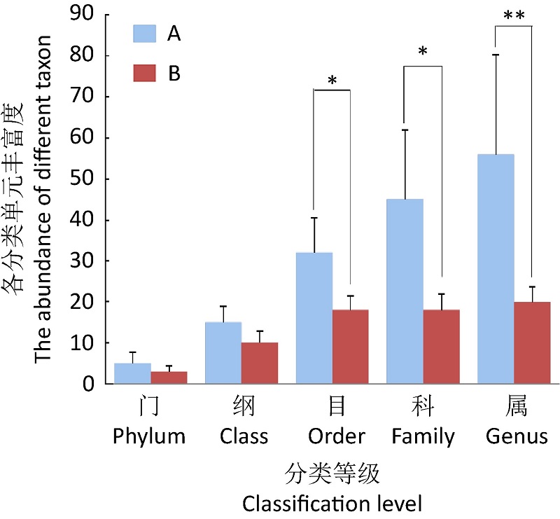
{{custom_ref.label}} |
{{custom_citation.content}}
{{custom_citation.annotation}}
|
/
〈 |
|
〉 |