响应面法优化六妹羊肚菌边角料中麦角硫因的提取工艺

张凤明, 甄梦玲, 赵硕, 田霞, 于富强

菌物学报 ›› 2025, Vol. 44 ›› Issue (3) : 240232.

PDF(876 KB)
中文  |  English
PDF(876 KB)
菌物学报 ›› 2025, Vol. 44 ›› Issue (3) : 240232. DOI: 10.13346/j.mycosystema.240232 CSTR: 32115.14.j.mycosystema.240232
研究论文

响应面法优化六妹羊肚菌边角料中麦角硫因的提取工艺

作者信息 +

Optimization of extraction process of ergothioneine from Morchella sextelata scraps by response surface methodology

Author information +
文章历史 +

摘要

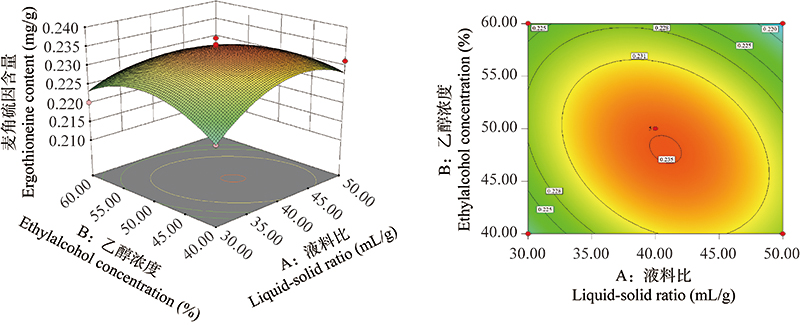
为提高六妹羊肚菌边角料中麦角硫因的得率,以麦角硫因得率为评价标准,分析了提取温度、乙醇浓度、液料比、提取时间等单因素对麦角硫因提取量的影响,在单因素实验的基础上通过响应面法对六妹羊肚菌边角料中麦角硫因的提取工艺条件进行了优化。单因素和响应面法得到的麦角硫因最佳提取工艺条件:液料比为40:1 (体积质量比)、乙醇浓度为50%、提取温度为60 ℃、提取时间为20 min。以该条件进行验证实验所得麦角硫因提取量为(0.236±0.000 887) mg/g,与预测值0.235 mg/g无显著差异(误差值0.426%),表明优化得到的工艺具有有效性和可靠性。本研究确定了六妹羊肚菌边角料中麦角硫因的最佳提取工艺,为六妹羊肚菌边角料的开发利用提供基础数据。

Abstract

The effect of extraction temperature, ethylalcohol concentration, liquid to material ratio and extraction time on the extraction yield of ergothioneine in M. sextelata scraps were studied by using the content of ergothioneine obtained as indicator, and the response surface methodology based on single factor experiment was applied to optimize the technological process of extraction. The results obtained by single factor experiment and response surface methodology indicated that liquid to material ratio of 40:1 (mL/g), ethylalcohol concentration of 50%, extraction temperature of 60 ℃, and extraction time of 20 min were the optimum extraction condition. Under this optimized condition, the extraction yield of ergothioneine was (0.236±0.000 887) mg/g, no significantly difference was found as compared with estimated value 0.235 mg/g (error value 0.426%), indicating that the optimized process was effective and reliable. This study provides basic data for utilization of M. sextelata scraps.

关键词

六妹羊肚菌 / 边角料 / 麦角硫因 / 响应面法 / 含量测定

Key words

Morchella sextelata / scrap / ergothioneine / response surface methodology / content determination

引用本文

导出引用
张凤明, 甄梦玲, 赵硕, 田霞, 于富强. 响应面法优化六妹羊肚菌边角料中麦角硫因的提取工艺[J]. 菌物学报, 2025, 44(3): 240232 https://doi.org/10.13346/j.mycosystema.240232
ZHANG Fengming, ZHEN Mengling, ZHAO Shuo, TIAN Xia, YU Fuqiang. Optimization of extraction process of ergothioneine from Morchella sextelata scraps by response surface methodology[J]. Mycosystema, 2025, 44(3): 240232 https://doi.org/10.13346/j.mycosystema.240232
银耳Tremella fuciformis Berk.属于担子菌门Basidiomycota银耳科Tremellaceae银耳属Tremella,是一种营养丰富、功能多元的食药用菌,作为药材和菜肴颇受消费者欢迎。据研究,银耳多糖(TPs)是银耳的主要功能成分,具有抗氧化、抗肿瘤、干预糖尿病、免疫调节和神经保护作用,同时,银耳多糖还能在皮肤抗衰老、光保护、伤口愈合和屏障保护方面发挥作用(Yang et al. 2019;Mineroff & Jagdeo 2023)。课题组前期从银耳子实体中分离纯化得到一种银耳多糖,能够干预葡聚糖硫酸钠诱导的小鼠结肠炎,同时能够激活Foxp3+T细胞,缓解肠道炎症,该银耳多糖还能显著增加肠道菌群多样性,恢复有益菌群相对丰度并结合菌群代谢物共同作用,达到干预结肠炎目的(Xu et al. 2021)。
肠道是人体最大的消化器官,也是免疫系统的重要组成部分,负责机体消化吸收营养物质、抵御外源抗原以及病原体侵染等,其中,肠道屏障的完整性、紧密性和稳定性是影响肠道作用的关键(Ohata et al. 2005;Kawai et al. 2021)。机械屏障是肠道屏障的支撑骨架,包括多种紧密连接蛋白形成的细胞间黏附复合结构,是机体化学屏障和生物屏障正常生理功能的结构基础,也是免疫屏障的首要防线,紧密连接蛋白受损会导致机体或者肠道炎症,是诱发各种疾病的重要机制(Suzuki 2020)。
研究表明紧密连接蛋白对干预消化道疾病和免疫疾病非常重要,保持紧密连接的完整性并提高其表达水平可有效干预相关疾病的发展(Miner-Williams & Moughan 2016;Zeisel et al. 2019;Tilg et al. 2020),脂多糖、炎症因子、高脂饮食通过不同机制都能导致肠道屏障受损(Lerner & Matthias 2015;Li et al. 2019;Ghezzal et al. 2020)。Ge et al. (2020)分离纯化的银耳多糖能够缓解脂多糖诱导的Caco-2细胞屏障损伤,目前尚没有银耳多糖能否缓解高脂饮食诱导的肠道紧密连接受损方面研究的报道。棕榈酸(palmitic acid, PAd)是人体内最常见的饱和脂肪酸,对人体不同器官有脂毒性,过量食用PAd会破坏紧密连接完整性,并触发炎症反应,导致肠道屏障功能障碍(Genser et al. 2018;Ghezzal et al. 2020)。
Ge et al. (2020)所分离得到的银耳多糖和课题组前期分离的银耳多糖在单糖组成、连接方式、取代基类型方面均不同(Xu et al. 2021),为了进一步探究课题组制得的银耳多糖是否是通过缓解肠道屏障损伤干预结肠炎,本研究以PAd刺激人结肠癌上皮细胞Caco-2建立紧密连接蛋白损伤模型,分析该银耳多糖处理后对Caco-2细胞活力、完整性、炎症因子、抗氧化性、紧密连接蛋白及其相关调控信号通路的效果,探究银耳多糖影响肠道屏障的作用机制。

1 材料与方法

1.1 材料

银耳购自四川裕德源生态农业科技有限公司;人克隆结肠癌Caco-2细胞(C1115,ATCC:HTB-37)购自上海WHELAB生物科学有限公司;棕榈酸购自西安鲲创科技发展有限公司;离子交换填料DEAE Sepharose、凝胶分离柱Sephacryl S-400HR购自GE公司;IL-1β、IL-6、TNF-α试剂盒购自江苏酶免实业有限公司;MDA、CAT、SOD试剂盒购自南京建成生物工程研究所;RIPA裂解液购自北京索莱宝生物科技有限公司;BCA蛋白浓度测定试剂盒购自碧云天生物科技有限公司。

1.2 方法

1.2.1 银耳多糖的提取纯化

根据课题组前期研究基础分离纯化得到一种银耳多糖(Xu et al. 2021),具体提取纯化方法如下:收集银耳子实体,以料液比1:20在沸水中浸提6 h,离心后取上清液3倍体积醇沉,采用Sevage法除蛋白(Miao et al. 2013),将除去蛋白的溶液透析后通过60 mm × 240 mm的DEAE柱进一步分离纯化,流速3.0 mL/min,分别用去离子水、50 mmol/L NaCl、150 mmol/L NaCl和1 mol/L NaCl进行洗脱,收集50 mmol/L NaCl洗脱液,再透析、冷冻干燥后得到分子量(Mw)为286.6 kDa的银耳多糖,单糖组成为甘露糖、核糖、鼠李糖、葡萄糖醛酸、葡萄糖、半乳糖、木糖、阿拉伯糖、岩藻糖,其组成比例为91 120.57、337.73、623.67、29 067.09、2 086.89、1 656.21、25 035.97、1 540.07、21 388.01 (Xu et al. 2021)。

1.2.2 细胞培养及处理

人克隆结肠癌Caco-2细胞采用10%胎牛血清加1%青链霉素混合液培养,置于37 ℃、5% CO2的细胞培养箱中培养。细胞贴壁生长,以1:3-1:5传代进行后续实验。取对数生长期细胞,加入不同浓度银耳多糖,最终浓度分别为25、50、100、200、400、600 μg/mL,每组设5个复孔,在培养箱中培养24 h后检测细胞活力。Caco-2细胞分化21 d之后,在上述细胞活力基础上进行加药处理,分别为:对照组(未处理组)、棕榈酸、棕榈酸+ 25 μg/mL TPs、棕榈酸+ 50 μg/mL TPs、棕榈酸+ 100 μg/mL TPs、棕榈酸+ 200 μg/mL TPs、棕榈酸+ 400 μg/mL TPs、棕榈酸+ 600 μg/mL TPs (棕榈酸作用浓度400 μmol/L)。置于37 ℃、5% CO2的细胞培养箱中培养。

1.2.3 MTT法测定细胞活力

1.2.2基础上,银耳多糖和棕榈酸处理细胞24 h后,每孔加入100 μL 0.5 mg/mL MTT溶液,在37 ℃、5% CO2的细胞培养箱中孵育4 h,除去MTT溶液并用PBS洗后每孔加入100 μL DMSO,振荡溶解10 min后酶标仪检测570 nm处吸光度,按照以下公式计算细胞存活率:
 细胞存活率 (%)=A1A0A2A0×100%
其中A0为细胞背景孔的吸光度;A1为药物处理孔的吸光度;A2为空白对照孔的吸光度。

1.2.4 跨膜电阻值TEER的测定

培养瓶中Caco-2细胞用0.25%胰蛋白酶消化后接种至transwell培养板中,在transwell板的上室和下室分别加入DMEM培养基,上室接种Caco-2细胞,使细胞浓度达到1×10⁵个/mL,第一周隔天更换细胞培养基,第二周开始每天换液直至21 d。Caco-2细胞分化21 d后分别添加PAd或PAd +不同剂量的TPs,在37 ℃、5% CO2的细胞培养箱中培养24 h,采用电阻表测量TEER值监测细胞单层完整性,根据以下公式计算TEER值(24孔transwell培养板的薄膜面积为0.33 cm2):
TEER(Ω∙cm2)=[TEER样品孔(Ω)-TEER空白孔(Ω)]×面积(cm2)

1.2.5 荧光实时定量逆转录聚合酶链式反应

采用TRIzol试剂并按照说明书方法提取细胞总RNA,用核酸测定仪测定RNA的浓度和纯度,将检测合格的样品用于反转录合成cDNA。以cDNA为模板,引物见表1,95 ℃反应30 s,一次;95 ℃反应5 s,退火,60 ℃反应20 s,循环40次,进行扩增。以GAPDH为内参,采用2-ΔΔt法进行分析。
表1 qRT-PCR引物联序列

Table 1 Primers’ sequences for qRT-PCR

Gene Forward primer (5ʹ→3ʹ) Reverse primer (3ʹ→5ʹ)
GAPDH CTCCTCCTGTTCGACAGTCA CGACCAAATCCGTTGACTCC
Claudin-1 CACCGTCTGTGTTTGAGCA CAAACCACCGCTTACAGATG
Occludin GACTATGTGGAAAGAGTTGAC ACCGCTGCTGTAACGAG
ZO-1 TTCACGCAGTTACGAGCAAG TTGGTGTTTGAAGGCAGAGC

1.2.6 免疫印迹检测

使用RIPA裂解液提取细胞中的总蛋白,用BCA蛋白浓度测定试剂盒检测提取蛋白浓度。首先进行SDS-PAGE分析,在电压90 V运行30 min,随后设置110 V电压运行1.5 h,再在90 V电压下转膜2 h,随后封闭、一抗孵育并漂洗、二抗孵育并漂洗、显影、拍照,使用Image J软件进行定量,检测Claudin-1、Occludin、ZO-1、TLR4、MyD88和p-NF-κB的相对蛋白表达量。

1.2.7 检测炎性因子含量和抗氧化性

参照1.2.6中的方法提取细胞中蛋白,采用ELISA试剂盒检测促炎因子IL-1β、IL-6、TNF-α的含量,参照试剂盒说明书进行操作:在空白孔、标准孔、待测样品孔中分别加样,用封板膜封板后37 ℃孵育30 min,洗涤96孔板并拍干,加入酶标试剂后温育、洗涤、加入显色剂后37 ℃避光显色10 min,加入终止液终止反应,在450 nm测量各孔吸光度。参照1.2.6中的方法提取细胞蛋白,采用试剂盒检测细胞蛋白的MDA、CAT、SOD值,具体操作方法参考试剂盒说明书。

2 结果与分析

2.1 银耳多糖和棕榈酸对Caco-2细胞的毒性作用

当Caco-2细胞分别与不同浓度的TPs孵育24 h后,细胞存活率均在98%以上(图1A),说明TPs对Caco-2细胞没有毒性;当Caco-2细胞分别与不同处理组孵育24 h后,细胞存活率均在96%以上(图1B),说明棕榈酸和银耳多糖共同作用对Caco-2细胞没有毒性,选择棕榈酸和银耳多糖共同作用下Caco-2细胞存活率最高(98%)的浓度:100、200、400 μg/mL TPs继续开展实验。
图1 棕榈酸和银耳多糖对Caco-2细胞活力和完整性的影响 A:不同浓度TPs对细胞活力影响;B:TPs和PAd对细胞活力影响;C:TPs和PAd对细胞完整性的影响. PAd组与CON组相比,#P<0.05,##P<0.01;Pad+TPs处理组与PAd组相比,*P<0.05,**P<0.01. 下同

Fig. 1 Effects of palmitic acid (PAd) and Tremella fuciformis polysaccharides (TPs) on cell viability and TEER value of Caco-2. A: Effects of TPs concentration on cell viability; B: Effects of TPs and PAd on cell viability; C: Effects of TPs and PAd on cell integrity. PAd vs. CON, #P<0.05 and ##P<0.01; PAd+TPs treatment group vs. PAd, *P<0.05 and **P<0.01. The same below.

Full size|PPT slide

2.2 银耳多糖对棕榈酸损伤Caco-2细胞完整性的影响

TEER值能够反映细胞单层完整性,400 μmol/L PAd能够一定程度破坏细胞单层完整性,加入不同浓度TPs后细胞单层完整性有所提升,其中400 μg/mL的TPs提升效果最好,达到 1 917.3 Ω∙cm2,是PAd组TEER值的2.31倍,其次是200 μg/mL TPs,达到PAd组TEER值的1.88倍(图1C)。说明银耳多糖能够缓解PAd造成的Caco-2细胞完整性损伤。

2.3 银耳多糖对Caco-2细胞抗氧化性的影响

SOD、CAT、MDA值可以反映Caco-2细胞氧化状态,氧化还原平衡对维持肠道稳态、调控肠道炎症有重要作用(Campbell & Colgan 2019)。经PAd处理后,抗氧化指标SOD和CAT降低、氧化指标MDA升高(图2),说明PAd增加了Caco-2细胞的氧化程度。经TPs处理后,细胞氧化程度降低。200和400 μg/mL TPs均能有效提升细胞抗氧化性能,且高剂量作用更明显。100、200和400 μg/mL TPs都能显著降低Caco-2细胞氧化程度,200 μg/mL TPs的作用最明显。
图2 银耳多糖对棕榈酸诱导的Caco-2细胞氧化性的影响 A:CAT水平;B:SOD水平;C:MDA水平

Fig. 2 Effects of TPs on PAd-induced oxidation of Caco-2 cells. A: CAT level; B: SOD level; C: MDA level.

Full size|PPT slide

2.4 银耳多糖对Caco-2细胞炎症因子表达的影响

PAd不仅能够影响Caco-2细胞完整性,也会诱导促炎因子IL-1β、TNF-α、IL-6表达。加入PAd后,Caco-2细胞产生的IL-1β、TNF-α、IL-6均显著上升。经TPs共培养后IL-1β、TNF-α、IL-6含量均显著下降,200和400 μg/mL TPs抑制促炎因子释放的效果更好,与剂量呈正相关(图3),说明一定浓度范围内,TPs浓度越高,抑制IL-1β、TNF-α、IL-6表达的作用越强。
图3 银耳多糖对棕榈酸诱导的Caco-2细胞炎症因子释放量的影响 A:IL-1β水平;B:IL-6水平;C:TNF-α水平

Fig. 3 Effects of TPs on the release of pro-inflammatory factor of Caco-2 cells. A: IL-1β level; B: IL-6 level; C: TNF-α level.

Full size|PPT slide

2.5 银耳多糖对棕榈酸损伤紧密连接蛋白的影响

上述结果表明PAd能够一定程度损伤肠道细胞完整性,从而造成肠道屏障损伤,而TPs具有一定的修复作用。为了进一步明确TPs对PAd诱导的紧密蛋白损伤的修复机制,检测主要的紧密连接蛋白Claudin-1、Occludin、ZO-1的mRNA和蛋白表达情况。qRT-PCR实验表明,PAd处理后Claudin-1、Occludin、ZO-1的mRNA表达水平分别显著降低至对照组的69.11%、57.56%、52.45%,而TPs处理后Claudin-1、Occludin、ZO-1的mRNA表达量明显回升,200和400 μg/mL TPs提升3种蛋白mRNA表达量的效果优于100 μg/mL TPs (图4)。进一步采用Western blot验证,Claudin-1、Occludin和ZO-1的蛋白表达趋势和mRNA一致,PAd显著下调3种蛋白表达量,经TPs处理后3种蛋白的表达量均有效提升(图5)。综合mRNA和蛋白表达结果,针对Claudin-1,提升效果最好的是400 μg/mL的TPs,针对Occludin和ZO-1,提升效果最好的是200 μg/mL TPs。
图4 银耳多糖对棕榈酸诱导的紧密连接蛋白mRNA表达量的影响 A:Claudin-1水平;B:Occludin水平;C:ZO-1水平

Fig. 4 Effect of TPs on PAd-induced tight junction protein mRNA expression. A: Claudin-1 level; B: Occludin level; C: ZO-1 level.

Full size|PPT slide

图5 银耳多糖对棕榈酸诱导的紧密连接蛋白表达量的影响 A:Western blot检测Claudin-1、Occludin、ZO-1的蛋白条带;B:Claudin-1、Occludin、ZO-1的相对表达量

Fig. 5 Effects of TPs on PAd-induced tight junction protein expression. A: Protein bands of Claudin-1, Occludin and ZO-1 were detected by Western blot. B: Relative expression of Claudin-1, Occludin, and ZO-1.

Full size|PPT slide

2.6 银耳多糖对Caco-2细胞炎症信号通路的影响

银耳多糖能够抑制棕榈酸诱导的Caco-2细胞促炎因子分泌,而炎症会导致肠道紧密连接蛋白受损(Li et al. 2019),因此,进一步检测经典的炎症通路TRL4/MyD88/NF-κB,探究银耳多糖是否通过影响TRL4/MyD88/NF-κB炎症信号通路来调控肠道屏障。PAd处理后TRL4、MyD88和p-NF-κB的表达量显著上调,TPs处理能有效抑制3种蛋白的表达,随着剂量升高抑制的效果越好(图6),说明TPs能够通过抑制TRL4/MyD88/NF-κB炎症信号通路下调促炎因子分泌。
图6 银耳多糖对TRL4/MyD88/NF-κB信号通路的影响 A:Western blot检测TLR4、MyD88、p-NF-κB的蛋白条带;B:TLR4、MyD88、p-NF-κB的相对表达量

Fig. 6 Effects of TPs on TRL4/MyD88/NF-κB signaling pathway. A: Protein bands of TLR4, MyD88, and p-NF-κB were detected by Western blot. B: Relative expression of TLR4, MyD88, and p-NF-κB.

Full size|PPT slide

3 讨论

肥胖症的流行在世界范围内不断蔓延,大大增加了其他相关慢性代谢疾病的全球负担,如2型糖尿病、非酒精性脂肪性肝病和心血管疾病等(James et al. 2018)。肥胖通常伴随着机体激素、炎症、脂质和葡萄糖水平紊乱的改变,这些代谢疾病都与肠道屏障息息相关(Zhang et al. 2024)。研究表明,多种来源的多糖能够维护肠道屏障进而干预代谢疾病。茯苓多糖可缓解由细胞焦亡驱动的肠道屏障破坏,从而减轻非酒精性脂肪性肝炎(Ye et al. 2022),黄芩多糖通过改善肠道屏障功能和调节肠道菌群改善葡聚糖硫酸钠诱导的溃疡性结肠炎(Cui et al. 2021),果胶多糖通过调节肠道菌群,增加肠道屏障功能和免疫增强活性,达到抑制肥胖、脂肪肝和炎症的效果(Lee et al. 2022)。
高脂饮食诱导的肥胖大鼠和小鼠肠道紧密连接蛋白Occludin、Claudin-1等蛋白的表达水平受到明显抑制(Suzuki & Hara 2010;Cremonini et al. 2019),作为食物中常见的饱和脂肪酸,摄入PAd的小鼠肠道紧密连接蛋白也明显减少或易位,从而影响肠道屏障功能(Xiao et al. 2017;Zhang et al. 2019;何亚伦等 2022)。细胞活力和完整性实验结果表明,暴露在PAd和TPs下不会对Caco-2细胞产生毒性作用,PAd能够损伤Caco-2细胞完整性,经TPs处理可以减轻PAd对细胞造成的损伤。
氧化还原平衡对维持肠道屏障至关重要,随着体内活性氧水平积累、内源性抗氧化物质消耗,以及氧化应激标志物水平增加,过量的活性氧导致氧化应激诱发炎症,分泌促炎因子(Ge et al. 2022;苏安祥等 2022)。经PAd处理后的Caco-2细胞抗氧化物SOD和CAT水平降低,脂质过氧化物MDA升高。由于硫氧还蛋白系统(TRX)、谷胱甘肽系统(GSH)以及核因子E2相关因子2 (NRF2)都参与调节细胞氧化还原稳态,目前尚不明确TPs是通过什么途径调控Caco-2细胞的氧化还原状态,可能是单一途径或者是多途径共同调控,因此,调控作用与剂量不成正比(Muri & Kopf 2020)。促炎因子IL-1β、TNF-α、IL-6分泌增加,说明PAd引发Caco-2细胞氧化应激反应并诱发了炎症,而TPs处理能有效缓解PAd由氧化应激诱发的炎症。促炎因子能够通过调控紧密连接蛋白的表达和组装,作用于紧密连接蛋白的功能。研究表明,Caco-2细胞与TNF-α和IFN-γ共同培养导致紧密连接蛋白ZO-1、Occludin、Claudin-1和Claudin-4重组,降低肠道屏障功能(Zolotarevsky et al. 2002)。Claudin-2是IL-22在肠上皮中作用的一个关键靶蛋白,IL-22能够诱导Claudin-2表达上调,进而提高肠道上皮屏障通透性(Ong et al. 2020)。经TPs处理能显著缓解PAd导致的紧密连接蛋白Claudin-1、Occludin和ZO-1表达量降低,说明PAd由氧化应激诱发的炎症影响紧密连接蛋白表达,而TPs能够通过提升抗氧化性能、抑制促炎因子分泌、提升紧密连接蛋白表达水平达到维护肠道屏障的作用。研究发现氢能够还原多余自由基调节氧化应激平衡,通过该方法调控TLR-4/MyD88/NF-κB信号通路最终保护肠道屏障(Li et al. 2024),NF-κB表达水平与肠上皮细胞凋亡息息相关,进而影响肠道屏障功能(Yao & Cadwell 2020;Yue et al. 2020)。因此,结合经典的TLR-4/MyD88/NF-κB信号通路,确认TPs能够通过抑制TRL4/MyD88/NF-κB炎症信号通路下调促炎因子分泌,进而保护肠道屏障。
课题组前期研究表明,TPs能够调控肠道菌群及菌群代谢物(Xu et al. 2021),而肠道菌群紊乱也会导致肠道屏障受损(雷棋怡等 2024),目前发现TPs可以干预PAd导致的肠道屏障受损,未来可进一步探究TPs改变的肠道菌群是否对肠道屏障产生影响,从而更全面地说明TPs对肠道屏障的作用。

4 结论

高剂量的TPs能够显著提升细胞完整性和抗氧化性,通过抑制TRL4/MyD88/NF-κB炎症信号通路抑制促炎因子分泌,提升紧密连接蛋白Claudin-1、Occludin和ZO-1表达,达到保护肠道屏障的作用。本文为揭示TPs维持肠道屏障的机制和扩展其应用范围提供了科学依据和思路。

作者贡献

董茜:实验、论文撰写;张仕林:实验、论文撰写;谢丽源:提供实验材料、验证;张谦:数据管理、软件作图、验证数据;舒雪琴:软件使用、作图;何晓兰:提供菌种和实验材料、修改论文;彭卫红:修改论文;许瀛引:论文构思、修改论文。

利益冲突

作者声明,该研究不存在任何潜在利益冲突的商业或财务关系。

参考文献

[1]
Bazela K, Solyga-Zurek A, Debowska R, Rogiewicz K, Bartnik E, Eris I, 2014. L-ergothioneine protects skin cells against UV-induced damage-a preliminary study. Cosmetics, 1(1): 51-60
[2]
Borodinal I, Kenny LC, McCarthy CM, Paramasivan K, Pretorius E, Roberts TJ, van der Hoek SA, Kell DB, 2022. The biology of ergothioneine, an antioxidant nutraceutical. Nutrition Research Reviews, 33(2): 190-217
[3]
Cao L, 2020. Effects of Morchella esculenta polysaccharide on anti-fatigue and anti-hypoxia in mice. Edible Fungi of China, 39(6): 40-42 (in Chinese)
[4]
Cheah IK, Halliwell B, 2021. Ergothioneine, recent developments. Redox Biology, 42: 101868
[5]
Cheah IK, Tang RM, Yew TS, Lim KH, Halliwell B, 2017. Administration of pure ergothioneine to healthy human subjects: uptake, metabolism, and effects on biomarkers of oxidative damage and inflammation. Antioxidants & Redox Signaling, 26(5): 193-206
[6]
Chen Q, Che CC, Yang SS, Ding PP, Si MR, Yang G, 2023. Anti-inflammatory effects of extracellular vesicles from Morchella on LPS-stimulated RAW264.7 cells via the ROS-mediated p 38 MAPK signaling pathway. Molecular and Cellular Biochemistry, 478(2): 317-327
[7]
Chen SY, Ho KJ, Hsieh YJ, Wang LT, Mau JL, 2012a. Contents of lovastatin, γ-aminobutyric acid and ergothioneine in mushroom fruiting bodies and mycelia. LWT-Food Science and Technology, 47(2): 274-278
[8]
Chen SY, Ho KJ, Liang CH, Tsai CH, Huang LY, Mau JL, 2012b. Preparation of culinary-medicinal king oyster mushroom Pleurotus eryngii-fermented products with high ergothioneine content and their taste quality. International Journal of Medicinal Mushrooms, 14(1): 85-93
[9]
Cohen N, Cohen J, Asatiani MD, Varshney VK, Yu HT, Yang YC, Li YH, Mau JL, Wasser SP, 2014. Chemical composition and nutritional and medicinal value of fruit bodies and submerged cultured mycelia of culinary- medicinal higher basidiomycetes mushroom. International Journal of Medicinal Mushrooms, 16(3): 273-291
[10]
Dai YC, Yang ZL, 2008. A revised checklist of medicinal fungi in China. Mycosystema, 27: 801-824 (in Chinese)
摘要
More and more people have recently payed their attention to studies or investigations on medicinal fungi in China. However, many nomenclatural inconsistencies were found in the Chinese reports. The publications on Chinese medicinal fungi were critically reviewed. As a result, 473 medicinal fungi from China were enumerated in the present checklist. All of the names were checked or revised in accordance with the contemporary taxonomy and the latest version of the International Code of Botanical Nomenclature (Vienna Code). The ‘out-of-date’ names, ill-names, and un-existed names and misapplied names in the previous reports were revised. The common synonyms were listed after their valid names. The main medicinal functions of each species, together with the original or important references, were provided.
[11]
Daniel LS, Peters KM, Borradaile NM, 2021. Mushroom-derived medicine? Preclinical studies suggest potential benefits of ergothioneine for cardiometabolic health. International Journal of Molecular Sciences, 22(6): 3246
[12]
Dubost NJ, Beelman RB, Peterson D, Royse DJ, 2005. Identification and quantification of ergothioneine in cultivated mushrooms by liquid chromatography-mass spectroscopy. International Journal of Medicinal Mushrooms, 8(3): 215-222
[13]
Dubost NJ, Beelman RB, Royse DJ, 2007. Influence of selected cultural factors and postharvest storage on ergothioneine content of common button mushroom Agaricus bisporus (J. Lge.) Imbach (Agaricomycetideae). International Journal of Medicinal Mushrooms, 9(2): 163-176
[14]
E HC, Feng LL, Zhao XY, Li XB, Zhao ZY, He XW, Zhang YM, 2022. Determination of L-ergothioneine in edible fungi by ultra performance liquid chromatography and confirmation by mass spectrometry. Chinese Journal of Analysis Laboratory, 41(5): 529-534 (in Chinese)
[15]
Fan SH, Ren JX, Zhang JH, Jin XD, Bai BQ, 2019. Extraction optimization of Morchella esculenta polysaccharides by response surface methodology and its antioxidant activity. Science and Techology of Food Industry, 40(6): 179-185, 192 (in Chinese)
[16]
Feng LL, E HC, Zhang YM, Li XB, Zhou CY, Zhao XY, Ren JL, 2021. Research progress on extraction, separation and detection of ergothionine in edible fungi. Acta Edulis Fungi, 28(1): 115-123 (in Chinese)
[17]
Fovet T, Guilhot C, Delobel P, Chopard A, Py G, Brioche T, 2022. Ergothioneine improves aerobic performance without any negative effect on early muscle recovery signaling in response to acute exercise. Frontiers in Physiology, 13: 834597
[18]
Gursoy N, Sarikurkcu C, Cengiz M, Solak MH, 2009. Antioxidant activities, metal contents, total phenolics and flavonoids of seven Morchella species. Food and Chemical Toxicology, 47(9): 2381-2388
[19]
He XY, Zhou ZY, Chen YY, Zhao JC, Li FH, Ming J, 2023. Bioactivity of ergothioneine and its application in food industry: a review. Food and Fermentation Industries, 49(10): 285-292 (in Chinese)
Ergothioneine (EGT) is one of the most representative bioactive ingredients in edible fungi. In addition to being abundant in the fruiting bodies of edible fungi, it can also be synthesized by a variety of microorganisms, including actinomycetes, fungi, certain yeasts, and archaea through various pathways. EGT has a variety of physiological activities such as antioxidant, anti-inflammatory, and immune-enhancing effects. In the food industry, EGT also shows a good color-protecting effect and food preservation function due to its free radical scavenging ability and anti-lipid peroxidation ability. In this paper, the sources, biological activities, and applications of EGT in the food industry were reviewed to provide references for further research and application of EGT in the future.
[20]
Hu JJ, Zhang YX, Wang Y, Mo YL, Wang J, 2019. Optimization of extraction process of ergothioneine from Flammulina velutipes by response surface methodology. Science and Techology of Food Industry, 40(9): 171-177 (in Chinese)
[21]
Huang Y, 2019. The studies on structure identification, bioactivity activity and the mechanism of anti-tumor activity of the Morehella esculenta (L.) Pers. polysaccharide (ME-X). MS Thesis, China West Normal University, Nanchong. 1-78 (in Chinese)
[22]
Ishimoto T, Nakamichi N, Hosotani H, Masuo Y, Sugiura T, Kato Y, 2014. Organic cation transporter-mediated ergothioneine uptake in mouse neural progenitor cells suppresses proliferation and promotes differentiation into neurons. PLoS One, 9(2): e89434
[23]
Ito T, Kato M, Tsuchida H, Harada E, Niwa T, Osawa T, 2011. Ergothioneine as an anti-oxidative/anti-inflammatory component in several edible mushroom. Food Science and Technology Research, 17(2): 103-110
[24]
Kalaras MD, Richie JP, Calcagnotto A, Beelman RB, 2017. Mushrooms: a rich source of the antioxidants ergothioneine and glutathione. Food Chemistry, 233: 429-433
While mushrooms are the highest dietary source for the unique sulfur-containing antioxidant ergothioneine, little is known regarding levels of the major biological antioxidant glutathione. Thus, our objectives were to determine and compare levels of glutathione, as well as ergothioneine, in different species of mushrooms. Glutathione levels varied >20-fold (0.11-2.41mg/gdw) with some varieties having higher levels than reported for other foods. Ergothioneine levels also varied widely (0.15-7.27mg/gdw) and were highly correlated with those of glutathione (r=0.62, P<0.001). Both antioxidants were more concentrated in pileus than stipe tissues in selected mushrooms species. Agaricus bisporus harvested during the third cropping flush contained higher levels of ergothioneine and glutathione compared to the first flush, possibly as a response to increased oxidative stress. This study demonstrated that certain mushroom species are high in glutathione and ergothioneine and should be considered an excellent dietary source of these important antioxidants.Copyright © 2017 Elsevier Ltd. All rights reserved.
[25]
Kitsanayanyong L, Ohshima T, 2022. Ergothioneine: a potential antioxidative and antimelanosis agent for food quality preservation. FEBS Letters, 596(10): 1330-1347
[26]
Ko HJ, Kim J, Ahn M, Kim JH, Lee GS, Shin T, 2021. Ergothioneine alleviates senescence of fibroblasts induced by UVB damage of keratinocytes via activation of the Nrf2/HO-1 pathway and HSP 70 in keratinocytes. Experimental Cell Research, 400(1): 112516
[27]
Koh SS, Ooi SCY, Lui NMY, Qiong C, Ho LTY, Cheah IKM, Halliwell B, Herr DR, Ong WY, 2021. Effect of ergothioneine on 7-ketocholesterol-induced endothelial injury. Neuromolecular Medicine, 23(1): 184-198
Ergothioneine (ET) is a naturally occurring antioxidant that is synthesized by non-yeast fungi and certain bacteria. ET is not synthesized by animals, including humans, but is avidly taken up from the diet, especially from mushrooms. In the current study, we elucidated the effect of ET on the hCMEC/D3 human brain endothelial cell line. Endothelial cells are exposed to high levels of the cholesterol oxidation product, 7-ketocholesterol (7KC), in patients with cardiovascular disease and diabetes, and this process is thought to mediate pathological inflammation. 7KC induces a dose-dependent loss of cell viability and an increase in apoptosis and necrosis in the endothelial cells. A relocalization of the tight junction proteins, zonula occludens-1 (ZO-1) and claudin-5, towards the nucleus of the cells was also observed. These effects were significantly attenuated by ET. In addition, 7KC induces marked increases in the mRNA expression of pro-inflammatory cytokines, IL-1β IL-6, IL-8, TNF-α and cyclooxygenase-2 (COX2), as well as COX2 enzymatic activity, and these were significantly reduced by ET. Moreover, the cytoprotective and anti-inflammatory effects of ET were significantly reduced by co-incubation with an inhibitor of the ET transporter, OCTN1 (VHCL). This shows that ET needs to enter the endothelial cells to have a protective effect and is unlikely to act via extracellular neutralizing of 7KC. The protective effect on inflammation in brain endothelial cells suggests that ET might be useful as a nutraceutical for the prevention or management of neurovascular diseases, such as stroke and vascular dementia. Moreover, the ability of ET to cross the blood-brain barrier could point to its usefulness in combatting 7KC that is produced in the CNS during neuroinflammation, e.g. after excitotoxicity, in chronic neurodegenerative diseases, and possibly COVID-19-related neurologic complications.
[28]
Kuang MT, Xu JY, Li JY, Yang L, Hou B, Zhao Q, Hu JM, 2022. Purification, structural characterization and immunomodulatory activities of a polysaccharide from the fruiting body of Morchella sextelata. International Journal of Biological Macromolecules, 213: 394-403
[29]
Lee SR, Roh HS, Lee S, Park HB, Jang TS, Ko YJ, Baek KH, Kim KH, 2018. Bioactivity-guided isolation and chemical characterization of antiproliferative constituents from morel mushroom (Morchella esculenta) in human lung adenocarcinoma cells. Journal of Functional Foods, 40: 249-260
[30]
Li F, Wang KH, Dong XB, Xu HD, 2022. Structure, conformation and immunomodulatory activity of a polysaccharide from Morchella sextelata. International Journal of Food Science and Technology, 57(7): 4628-4637
[31]
Li JL, Liu YT, Zong S, Zhang JQ, Ye M, 2020. In vitro hyperglycemic and hypolipidemic activity of Morchella esculenta extra-cellular polysaccharides. Food Research and Development, 41(16): 39-45 (in Chinese)
[32]
Li YH, 2016. Study on processes of extraction and purification of ergothioneine from Pleurotus eryngii and its antioxidant activity. MS Thesis, South China Agricultural University, Guangzhou. 1-73 (in Chinese)
[33]
Li YL, Zhang YF, Liu Z, Liu D, Chi RA, 2015. Optimization of microwave assisted extraction of ginsenosides and polysaccharide by respone surface methodology. China Medical Herald, 12(16): 42-47 (in Chinese)
[34]
Martinez-Medina GA, Chávez-González ML, Verma DK, Prado-Barragán LA, Martínez-Hernández JL, Flores-Gallegos AC, Thakur M, Srivastav PP, Aguilar CN, 2021. Bio-funcional components in mushrooms, a health opportunity: ergothionine and huitlacohe as recent trends. Journal of Functional Foods, 77: 104326
[35]
Mo YL, Li YH, Wang Y, Hu JJ, Zhang YX, Wang J, 2020. Optimization of ultrasonic combined microwave-assisted extraction of ergothioneine in Pleurotus eryngii by response surface methodology. Science and Techology of Food Industry, 41(1): 143-149 (in Chinese)
[36]
Mukaidaisi MHMT, 2023. Isolation and purification of ergosthioneine from Phlebopus portentosus and transcriptomic study on its inhibition of melanin production in B16F10 cells. MS Thesis, Zhongkai University of Agriculture and Engineering, Guangzhou. 1-69 (in Chinese)
[37]
Nitha B, Fijesh PV, Janardhanan KK, 2013. Hepatoprotective activity of cultured mycelium of morel mushroom, Morchella esculenta. Experimental and Toxicologic Pathology, 65(1-2): 105-112
The hepatoprotective activity of cultured mycelium of morel mushroom Morchella esculenta against CCl(4) and ethanol induced chronic hepatotoxicity was investigated. Hepatotoxicity was induced by challenging the animals with CCl(4) (1:5, v/v, 3.75 ml/kg body weight, i.p., 30 doses) and ethanol (36%, v/v, 6 ml/animal, p.o., 35 doses) and the extract was administered at two concentrations (250 and 500 mg/kg body weight). Hepatoprotection was evaluated by determining the activities of liver function marker enzymes and antioxidant status of liver and also by histopathological observations of liver tissue. Administration of both ethanol and CCl(4) elevated the levels of liver function enzymes, GOT, GPT and ALP in serum drastically. The treatment with the extract decreased the elevated serum GOT, GPT and ALP activities in a dose dependent manner. The extract also restored the depleted levels of antioxidants in liver consequent to CCl(4) and ethanol challenge. The results indicated that aqueous-ethanolic extract of M. esculenta mycelium possessed significant hepatoprotective activity. The conclusion is also supported by the biochemical determinations and histopathological observations. The findings thus suggest the potential therapeutic use of morel mushroom mycelium as a novel hepatoprotective agent.Copyright © 2011 Elsevier GmbH. All rights reserved.
[38]
Nitha B, Janardhanan KK, 2008. Aqueous-ethanolic extract of morel mushroom mycelium Morchella esculenta, protects cisplatin and gentamicin induced nephrotoxicity in mice. Food and Chemical Toxicology, 46(9): 3193-3199
Morchella esculenta (L) Pers. is an excellently edible and delicious morel mushroom found growing in the temperate forests. The mycelium of this mushroom is widely used as a flavouring agent. The current investigation was undertaken to explore the protective effect of the aqueous-ethanol extract of cultured mycelium of M. esculenta against cisplatin and gentamicin induced acute renal toxicity in Swiss albino mice. Cisplatin and gentamicin when administered induced a marked renal failure, characterized by a significant increase in serum urea and creatinine concentrations. Treatment with the extract at 250 and 500mg/kg body weight decreased the cisplatin and gentamicin induced increase in serum creatinine and urea levels. Treatment with the extract also restored the depleted antioxidant defense system. The decreased activity of superoxide dismutase (SOD), catalase (CAT), glutathione peroxidase (GPx), and reduced glutathione (GSH) in the kidneys consequent to cisplatin and gentamicin administration was significantly elevated. The enhanced renal antioxidant defense system also prevented the tissue lipid peroxidation. The experimental results suggest that aqueous-ethanol extract of morel mushroom, M. esculenta mycelium protected cisplatin and gentamicin induced nephrotoxicity possibly by enhancing renal antioxidant system. The findings thus suggest the potential therapeutic use of morel mushroom mycelium as a novel nephroprotective agent.
[39]
Pan HY, Guo LQ, Lin JF, 2019. Recent advances in understanding the in vivo distribution and metabolism of ergothioneine and its roles in disease prevention. Food Science, 40(23): 334-340 (in Chinese)
[40]
Paul BD, Snyder SH, 2009. The unusual amino acid L-ergothioneine is a physiologic cytoprotectant. Cell Death and Differentiation, 17(7): 1134-1140
[41]
Ramya H, Ravikumar KS, Fathimathu Z, Janardhanan KK, Ajith TA, Shah MA, Farooq R, Reshi ZA, 2022. Morel mushroom, Morchella from Kashmir Himalaya: a potential source of therapeutically useful bioactives that possess free radical scavenging, anti-inflammatory, and arthritic edema-inhibiting activities. Drug and Chemical Toxicology, 45(5): 2014-2023
[42]
Smith E, Ottosson F, Hellstrand S, Ericson U, Orho-Melander M, Fernandez C, Melander O, 2020. Ergothioneine is associated with reduced mortality and decreased risk of cardiovascular disease. Heart, 106(9): 691-697
We recently identified a health conscious food pattern (HCFP) associated with reduced risk of cardiometabolic disease. However, the molecular events linking the healthy food pattern to reduced risk of cardiometabolic disease are unknown. Our aim was to identify plasma metabolites associated with the HCFP and test if such metabolites predict cardiometabolic disease and mortality.Using liquid-chromatography mass-spectrometry, 112 plasma metabolites were measured in 3236 participants without cardiovascular disease (CVD) and diabetes mellitus from the population-based Malmö Diet and Cancer study. Metabolites associated with the HCFP were identified using multivariable adjusted linear regressions followed by Bonferroni correction. The healthy dietary biomarkers were subsequently related to risk of cardiometabolic disease and mortality during long-term follow-up with multivariable adjusted Cox proportional hazards models.During a median follow-up time of 21.4 years, 603 participants developed CVD, 362 developed diabetes mellitus and 843 participants died. Five healthy dietary biomarkers were associated with the HCFP at baseline (p<0.0004) and four predicted at least one of the studied end points (p<0.05). Ergothioneine was the metabolite most strongly connected to the HCFP and was associated with a lower risk of coronary disease (HR per 1 SD increment of ergothioneine, HR=0.85, p=0.01), cardiovascular mortality (HR=0.79, p=0.002) and overall mortality (HR=0.86, p=4e-5).We identified that higher ergothioneine was an independent marker of lower risk of cardiometabolic disease and mortality, which potentially can be induced by a specific healthy dietary intake.© Author(s) (or their employer(s)) 2020. Re-use permitted under CC BY-NC. No commercial re-use. See rights and permissions. Published by BMJ.
[43]
Tang Y, Chen JL, Li FH, Yang YX, Wu SR, Ming J, 2019. Antioxidant and antiproliferative activities of modified polysaccharides originally isolated from Morchella angusticepes Peck. Journal of Food Science, 84(3): 448-456
Polysaccharides extracted from Morchella angusticepes Peck (PMEP) were chemically modified to obtain sulfated, carboxymethylated, and acetyled derivatives. Results showed that the acetyled derivatives with the degree of substitution (0.40 ± 0.07) exerted higher antioxidant ability than native polysaccharides, demonstrating inhibitory effects on growth of human hepatoma cells (EC  = 0.710 ± 0.002 mg/mL) and human colon cancer cells (EC = 1.229 ± 0.008 mg/mL). It indicated that the acetylation was a favorable way to enhance the bioactivities of PMEP. PRACTICAL APPLICATION: Polysaccharides extracted from Morchella angusticepes Peck (PMEP) have many health-promoting properties. Chemical modifications could improve the bioactivities of polysaccharides. We demonstrated that acetylation enhanced the cellular antioxidant and antiproliferative activities of PMEP. The results support further research that explores clinical utility, and may justify ex vivo and in vivo designs toward that end.© 2019 Institute of Food Technologists®.
[44]
Tanret C, 1909. The new base drawn from rye ergot, ergothioneine. Comptes Rendus Hebdomadaires des Séances de l'Académie des Sciences, 149: 222-224
[45]
Tepwong P, Giri A, Sasaki F, Fukui R, Ohshima TM, 2012. Mycobial enhancement of ergothioneine by submerged cultivation of edible mushroom mycelia and its application as an antioxidative compound. Food Chemistry, 131(1): 247-258
[46]
Tian JF, Shang YH, Xiao ZN, 2024. Research progress on nutrient composition, function and processing of morels. Science and Techology of Food Industry, 45(9): 419-428 (in Chinese)
[47]
Tietel Z, Masaphy S, 2018. True morels (Morchella)-nutritional and phytochemical composition, health benefits and flavor: a review. Critical Reviews in Food Science and Nutrition, 58(11): 1888-1901
Morels are edible mushrooms appreciated worldwide for their savory flavor. Morels have been in use in traditional medicine for centuries, due to their health-related benefits, and current research demonstrated their anti-oxidative and anti-inflammatory bioactivities, in addition to immunostimulatory and anti-tumor properties. In spite of the high demand for morels and their increasing economic importance, their cultivation is limited, and they are either used as wild harvested or fermented in culture, for consumption as a functional food and for food-flavoring. Morel's health benefits were attributed mainly to polysaccharides as the active compounds, and to various phytochemicals, mainly phenolic compounds, tocopherols, ascorbic acid and vitamin D. Morel's nutritional composition was reported, including sugar, amino acid, fatty and organic acid and mineral profile. Information regarding Morel's flavor is limited, and while some of their taste attributes have been described, including the role of umami taste, details about their volatile aroma profile are scarce, and it was reported to include eight carbon volatiles, the main aroma volatiles typical to most mushrooms. To the best of our knowledge, this is the first review presenting morels' nutritional and phytochemical composition, health benefits and flavor, and we will review the available information in current literature regarding these aspects in light of morels phenotypic plasticity.
[48]
Tucker RAJ, Cheah IK, Halliwell B, 2019. Specificity of the ergothioneine transporter natively expressed in HeLa cells. Biochemical and Biophysical Research Communications, 513(1): 22-27
Ergothioneine is a biologically important compound that has been shown to be transported by the organic cation transporter novel type 1 (OCTN1). Following this discovery, a variety of alternate functions for OCTN1 have been suggested including an integral function in the extra-neuronal cholinergic system. The present study reaffirms the primacy of ergothioneine over these alternate substrates using natively expressed OCTN1 in HeLa cells. Besides the general transport inhibitors, quinidine, verapamil and pyrilamine no other putative substrate inhibited ergothioneine transport significantly, with only a slight inhibition demonstrated by carnitine. Even compounds structurally similar to ergothioneine failed to inhibit ergothioneine uptake, suggesting high selectivity of OCTN1. Ergothioneine was found to be avidly accumulated even at low concentrations (300 nM) by HeLa cells.Copyright © 2019 Elsevier Inc. All rights reserved.
[49]
Valachova K, Svik K, Biro C, Collins MN, Jurcik R, Ondruska L, Soltes L, 2020. Impact of ergothioneine, hercynine, and histidine on oxidative degradation of hyaluronan and wound healing. Polymers, 13(1): 95
[50]
Wan AL, 2019. Study of protective effects of ergothioneine in five boletus on liver injury induced by carbon tetrachloride in mice. MS Thesis, Kunming University of Science and Technology, Kunming. 1-86 (in Chinese)
[51]
Wang DD, Yin ZQ, Ma LK, Han L, Chen Y, Pan WJ, Gong K, Gao YY, Yang XX, Chen YL, Han JH, Duan Y, 2021. Polysaccharide MCP extracted from Morchella esculenta reduces atherosclerosis in LDLR-deficient mice. Food & Function, 12(11): 4842-4854
[52]
Wang YH, Yan LL, Li JR, Zheng GY, 2023. Optimizing the extraction of ergothioneine from Stropharia rugosoannulata by response surface methodology and evaluating the stability of in vitro antioxidant capacity. Journal of Food Safety and Quality, 14(16): 293-302 (in Chinese)
[53]
Wu F, Zhou LW, Yang ZL, Bau T, Li TH, Dai YC, 2019. Resource diversity of Chinese macrofungi: edible, medicinal and poisonous species. Fungal Diversity, 98: 1-76
[54]
Xu HY, Xie ZL, Meng Q, Ma DJ, Sun LZ, 2022. Oxidative damage and antioxidant defenses of Morchella spongiola in response to Cd stress. Mycosystema, 41(4): 668-679 (in Chinese)
[55]
Xue L, 2014. Inhibitory effects of extracellular polysaccharides extracted from Morchella esculenta on S180sarcoma. Journal of Shanxi College of Traditional Chinese Medicine, 15(2): 27-29 (in Chinese)
[56]
Yang H, Yan MM, 2012. Extraction of jujube polysaccharides with pectinase. Journal of Shaanxi University of Science & Technology, 30(5): 62-66 (in Chinese)
[57]
Yoshida S, Shime H, Funami K, Takaki H, Matsumoto M, Kasahara M, Seya T, 2017. The anti-oxidant ergothioneine augments the immunomodulatory function of TLR agonistsby direct action on macrophages. PLoS One, 12(1): e0169360
[58]
Zhang C, Zhao YM, Bai SF, Liu DL, 2013. HPLC determination of ergothioneine in mushrooms of different species. Science and Technology of Food Industry, 34(23): 307-310 (in Chinese)
[59]
Zhao YM, Lei ZD, Liu CH, Liu DL, 2016. Determination of ergothioneine in multiple species of fungi by HPLC. Food Research and Development, 37(2): 117-119 (in Chinese)
[60]
曹亮, 2020. 羊肚菌多糖对小鼠抗运动疲劳及耐缺氧的影响. 中国食用菌, 39(6): 40-42
[61]
戴玉成, 杨祝良, 2008. 中国药用真菌名录及部分名称的修订. 菌物学报, 27: 801-824
[62]
鄂恒超, 冯路路, 赵晓燕, 李晓贝, 赵志勇, 何香伟, 张艳梅, 2022. 超高效液相色谱法测定食用菌中L-麦角硫因及质谱确证. 分析试验室, 41(5): 529-534
[63]
范三红, 任嘉兴, 张锦华, 金晓弟, 白宝清, 2019. 响应面优化羊肚菌多糖提取工艺及抗氧化性. 食品工业科技, 40(6): 179-185, 192
[64]
冯路路, 鄂恒超, 张艳梅, 李晓贝, 周昌艳, 赵晓燕, 任佳丽, 2021. 食用菌中麦角硫因提取分离和检测方法研究进展. 食用菌学报, 28(1): 115-123
[65]
何鑫怡, 周子艺, 陈媛媛, 赵吉春, 李富华, 明建, 2023. 麦角硫因生物活性及其在食品工业中的应用. 食品与发酵工业, 49(10): 285-292
摘要
麦角硫因(ergothioneine, EGT)是食用菌最具代表性的生物活性成分之一。除了广泛存在于食用菌子实体,还可以由多种微生物,包括放线菌、真菌、某些酵母菌和古细菌通过需氧或厌氧途径合成。EGT具有抗氧化、抗炎和提高免疫等多种生理活性。在食品工业中,基于其自由基清除能力和抗脂质过氧化能力,EGT还表现出了良好的护色效果和食品保藏功能。论文综述了EGT的来源、生物活性和在食品工业中的应用,旨在为EGT今后的进一步研究和应用提供参考。
[66]
胡晶晶, 张怡馨, 王艳, 莫宇丽, 王杰, 2019. 响应面法优化金针菇麦角硫因的提取工艺. 食品工业科技, 40(9): 171-177
[67]
黄瑶, 2019. 羊肚菌多糖(ME-X)结构鉴定、生物活性及抗肿瘤机制的研究. 西华师范大学硕士论文, 南充. 1-78
[68]
李井雷, 刘玉婷, 宗帅, 张家奇, 叶明, 2020. 羊肚菌胞外多糖体外降血糖降血脂活性研究. 食品研究与开发, 41(16): 39-45
[69]
李亚欢, 2016. 杏鲍菇中麦角硫因的提取纯化和抗氧化活性研究. 华南农业大学硕士论文, 广州. 1-73
[70]
李娅丽, 张越非, 刘哲, 刘迪, 池汝安, 2015. 响应曲面法优化人参皂苷及多糖微波辅助提取工艺. 中国医药导报, 12(16): 42-47
[71]
莫宇丽, 李亚欢, 王艳, 胡晶晶, 张怡馨, 王杰, 2020. 响应面法优化超声微波联合提取杏鲍菇中麦角硫因工艺. 食品工业科技, 41(1): 143-149
[72]
木开代斯·买合木提, 2023. 黑牛肝菌麦角硫因的分离纯化及抑制B16F10细胞黑色素生成的转录组学研究. 仲恺农业工程学院硕士论文, 广州. 1-69
[73]
潘虹余, 郭丽琼, 林俊芳, 2019. 麦角硫因机体内的分布与代谢和其在疾病中的作用研究进展. 食品科学, 40(23): 334-340
麦角硫因是一种稀有的天然氨基酸类强抗氧化剂,由放线菌、蓝细菌、部分真菌(例如链霉菌、分枝杆菌等)和蘑菇等微生物合成。动物(包括人类)和植物自身不能在体内合成麦角硫因,植物可通过根系从土壤中吸收微生物合成的麦角硫因,而动物可从食物中吸收和积累麦角硫因。人体通过有机阳离子转运蛋白1型从食物中吸收麦角硫因,其广泛分布于人体的细胞和组织中。已有大量研究报道了麦角硫因的合成、自然界中的分布、化学性质、生理功能和其作为抗氧化剂的特性。尽管研究证明麦角硫因在体外具有明显的抗氧化活性和细胞保护作用,但对其在体内的生理功能研究仍有限。本文旨在综述麦角硫因的独特性质、生物合成以及预防疾病的潜力。
[74]
田金凤, 尚远宏, 肖宗妮, 2024. 羊肚菌的营养成分、功能和加工的研究进展. 食品工业科技, 45(9): 419-428
[75]
万安露, 2019. 五种牛肝菌中麦角硫因对四氯化碳致小鼠肝损伤的保护作用. 昆明理工大学硕士论文, 昆明. 1-86
[76]
王艳辉, 闫林林, 李俊仁, 郑光耀, 2023. 响应面法优化大球盖菇麦角硫因的提取工艺及其体外抗氧化能力稳定性的考察. 食品安全质量检测学报, 14(16): 293-302
[77]
徐鸿雁, 谢占玲, 孟清, 马德娟, 孙灵芝, 2022. 镉胁迫小海绵羊肚菌氧化损伤及其抗氧化防御. 菌物学报, 41(4): 668-679
羊肚菌Morchella是全球广泛分布的食药用真菌,重金属镉(Cd)在羊肚菌中的积累受到越来越多的关注。然而,羊肚菌镉积累的机理尚不清楚。本研究通过在0-5.0 mg/L Cd浓度环境中培养小海绵羊肚菌Morchella spongiola,测定Cd胁迫下其菌丝生长速率、丙二醛(MDA)、过氧化氢(H<sub>2</sub>O<sub>2</sub>)、超氧化物歧化酶(SOD)、过氧化物酶(POD)、过氧化氢酶(CAT)、谷胱甘肽过氧化物酶(GSH-Px)、谷胱甘肽(GSH)、抗坏血酸(ASA)及细胞Cd积累量等生理生化指标,旨在明晰小海绵羊肚菌响应Cd毒害的抗氧化防御响应机理。结果表明随着Cd浓度的增加,小海绵羊肚菌菌丝生长呈现出&#x0201c;升-降-升-降&#x0201d;的双峰响应变化,其中0.15、0.90和1.50 mg/L为菌丝生长Cd浓度关键拐点。Cd胁迫导致的氧化损伤与其初始浓度呈现正相关,胁迫3 d时MDA和H<sub>2</sub>O<sub>2</sub>含量显示出较大提升,5.0 mg/L处理组MDA和H<sub>2</sub>O<sub>2</sub>含量比对照组分别高出5.80倍和6.08倍。胁迫浓度、胁迫时间对抗氧化系统的影响各异,SOD、POD活性随浓度增大而逐渐升高,在1.5-5.0 mg/L浓度范围内,SOD和POD酶活性分别增加了1.96倍和2.15倍;CAT和GSH-Px酶活在0.15 mg/L时达到最大值,之后增加Cd浓度酶活性被抑制;GSH和ASA含量在Cd胁迫初期呈浓度依赖性增加,胁迫5 d后表现二者含量先升高后降低。此外,小海绵羊肚菌胞外Cd积累量随着外源Cd浓度的提高而增加,而胞内Cd含量在浓度为1.5-5.0 mg/L范围内无明显增加。本研究首次系统研究了小海绵羊肚菌响应Cd胁迫浓度、胁迫时间的动态趋势与内在变化规律,不仅为羊肚菌与重金属相互作用关系研究奠定基础,也为羊肚菌用于Cd污染废水生物修复提供了理论依据。
[78]
薛莉, 2014. 羊肚菌胞外粗多糖对S180肉瘤小鼠的抑制实验. 山西中医学院学报, 15(2): 27-29
[79]
杨辉, 闫明明, 2012. 果胶酶提取大枣多糖的工艺研究. 陕西科技大学学报, 30(5): 62-66
[80]
张翠, 赵艳敏, 白淑芳, 刘岱琳, 2013. HPLC法测定不同品种蘑菇中麦角硫因的含量. 食品工业科技, 34(23): 307-310
[81]
赵艳敏, 雷智东, 刘成航, 刘岱琳, 2016. 高效液相色谱法测定多种真菌中麦角硫因含量. 食品研究与开发, 37(2): 117-119

基金

云南省重大科技专项计划(202202AE090001)
云南省博士后定向资助项目(E23174K2)
云南省博士后科研基金(E2313442)
PDF(876 KB)

Accesses

Citation

Detail

段落导航
相关文章

/