银耳多糖对肠道屏障的影响及作用机制

董茜, 张仕林, 谢丽源, 张谦, 舒雪琴, 何晓兰, 彭卫红, 许瀛引

菌物学报 ›› 2025, Vol. 44 ›› Issue (3) : 240251.

PDF(782 KB)
中文  |  English
PDF(782 KB)
菌物学报 ›› 2025, Vol. 44 ›› Issue (3) : 240251. DOI: 10.13346/j.mycosystema.240251 CSTR: 32115.14.j.mycosystema.240251
研究论文

银耳多糖对肠道屏障的影响及作用机制

作者信息 +

Effects of Tremella fuciformis polysaccharides on intestinal barrier and its functional mechanism

Author information +
文章历史 +

摘要

以棕榈酸刺激人结肠癌上皮细胞Caco-2构建高脂膳食模式下的紧密连接蛋白损伤模型,研究银耳多糖对肠道屏障的影响及作用机制。通过检测细胞活力、细胞完整性、氧化还原水平、炎症因子含量、紧密连接基因和蛋白表达量,以及炎症信号通路表达水平,发现400 μg/mL TPs银耳多糖对Caco-2细胞无毒性,能够提升细胞SOD和CAT含量并降低MDA含量,抑制促炎因子IL-1β、TNF-α、IL-6释放,提升紧密连接蛋白Claudin-1、Occludin和ZO-1的mRNA和蛋白表达水平,通过抑制TRL4/MyD88/NF-κB炎症信号通路下调促炎因子分泌。银耳多糖通过抗氧化、抑制炎症因子分泌、提升紧密连接蛋白表达参与维护肠道屏障。

Abstract

Palmitic acid was used to stimulate human colon cancer epithelial cells Caco-2 to construct a tight-junction protein injury model under high fat diet for studying the effect of Tremella fuciformis polysaccharides on intestinal barrier and its functional mechanism. By measuring cell viability, cell integrity, redox level, inflammatory factor content, tight junction gene, protein expression level, and inflammatory signaling pathway expression level, it was found that 400 μg/mL T. fuciformis polysaccharides was non-toxic to Caco-2 cells, and could increase SOD and CAT content and decrease MDA content, inhibit the release of pro-inflammatory factors IL-1β, TNF-α and IL-6, enhance the mRNA and protein expression levels of tight junction proteins Claudin-1, Occludin and ZO-1, and down-regulate the secretion of pro-inflammatory factors by inhibiting the inflammatory signaling pathway of TRL4/MyD88/NF-κB. T. fuciformis polysaccharides maintained the intestinal barrier by means of anti-oxidation, inhibiting the secretion of inflammatory factors and elevating the expression of tight junction protein.

关键词

银耳多糖 / 棕榈酸 / 肠道炎症 / 肠道屏障

Key words

Tremella fuciformis polysaccharides / palmitic acid / inflammatory bowel disease / gut barrier

引用本文

导出引用
董茜, 张仕林, 谢丽源, 张谦, 舒雪琴, 何晓兰, 彭卫红, 许瀛引. 银耳多糖对肠道屏障的影响及作用机制[J]. 菌物学报, 2025, 44(3): 240251 https://doi.org/10.13346/j.mycosystema.240251
DONG Qian, ZHANG Shilin, XIE Liyuan, ZHANG Qian, SHU Xueqin, HE Xiaolan, PENG Weihong, XU Yingyin. Effects of Tremella fuciformis polysaccharides on intestinal barrier and its functional mechanism[J]. Mycosystema, 2025, 44(3): 240251 https://doi.org/10.13346/j.mycosystema.240251
银耳Tremella fuciformis Berk.属于担子菌门Basidiomycota银耳科Tremellaceae银耳属Tremella,是一种营养丰富、功能多元的食药用菌,作为药材和菜肴颇受消费者欢迎。据研究,银耳多糖(TPs)是银耳的主要功能成分,具有抗氧化、抗肿瘤、干预糖尿病、免疫调节和神经保护作用,同时,银耳多糖还能在皮肤抗衰老、光保护、伤口愈合和屏障保护方面发挥作用(Yang et al. 2019;Mineroff & Jagdeo 2023)。课题组前期从银耳子实体中分离纯化得到一种银耳多糖,能够干预葡聚糖硫酸钠诱导的小鼠结肠炎,同时能够激活Foxp3+T细胞,缓解肠道炎症,该银耳多糖还能显著增加肠道菌群多样性,恢复有益菌群相对丰度并结合菌群代谢物共同作用,达到干预结肠炎目的(Xu et al. 2021)。
肠道是人体最大的消化器官,也是免疫系统的重要组成部分,负责机体消化吸收营养物质、抵御外源抗原以及病原体侵染等,其中,肠道屏障的完整性、紧密性和稳定性是影响肠道作用的关键(Ohata et al. 2005;Kawai et al. 2021)。机械屏障是肠道屏障的支撑骨架,包括多种紧密连接蛋白形成的细胞间黏附复合结构,是机体化学屏障和生物屏障正常生理功能的结构基础,也是免疫屏障的首要防线,紧密连接蛋白受损会导致机体或者肠道炎症,是诱发各种疾病的重要机制(Suzuki 2020)。
研究表明紧密连接蛋白对干预消化道疾病和免疫疾病非常重要,保持紧密连接的完整性并提高其表达水平可有效干预相关疾病的发展(Miner-Williams & Moughan 2016;Zeisel et al. 2019;Tilg et al. 2020),脂多糖、炎症因子、高脂饮食通过不同机制都能导致肠道屏障受损(Lerner & Matthias 2015;Li et al. 2019;Ghezzal et al. 2020)。Ge et al. (2020)分离纯化的银耳多糖能够缓解脂多糖诱导的Caco-2细胞屏障损伤,目前尚没有银耳多糖能否缓解高脂饮食诱导的肠道紧密连接受损方面研究的报道。棕榈酸(palmitic acid, PAd)是人体内最常见的饱和脂肪酸,对人体不同器官有脂毒性,过量食用PAd会破坏紧密连接完整性,并触发炎症反应,导致肠道屏障功能障碍(Genser et al. 2018;Ghezzal et al. 2020)。
Ge et al. (2020)所分离得到的银耳多糖和课题组前期分离的银耳多糖在单糖组成、连接方式、取代基类型方面均不同(Xu et al. 2021),为了进一步探究课题组制得的银耳多糖是否是通过缓解肠道屏障损伤干预结肠炎,本研究以PAd刺激人结肠癌上皮细胞Caco-2建立紧密连接蛋白损伤模型,分析该银耳多糖处理后对Caco-2细胞活力、完整性、炎症因子、抗氧化性、紧密连接蛋白及其相关调控信号通路的效果,探究银耳多糖影响肠道屏障的作用机制。

1 材料与方法

1.1 材料

银耳购自四川裕德源生态农业科技有限公司;人克隆结肠癌Caco-2细胞(C1115,ATCC:HTB-37)购自上海WHELAB生物科学有限公司;棕榈酸购自西安鲲创科技发展有限公司;离子交换填料DEAE Sepharose、凝胶分离柱Sephacryl S-400HR购自GE公司;IL-1β、IL-6、TNF-α试剂盒购自江苏酶免实业有限公司;MDA、CAT、SOD试剂盒购自南京建成生物工程研究所;RIPA裂解液购自北京索莱宝生物科技有限公司;BCA蛋白浓度测定试剂盒购自碧云天生物科技有限公司。

1.2 方法

1.2.1 银耳多糖的提取纯化

根据课题组前期研究基础分离纯化得到一种银耳多糖(Xu et al. 2021),具体提取纯化方法如下:收集银耳子实体,以料液比1:20在沸水中浸提6 h,离心后取上清液3倍体积醇沉,采用Sevage法除蛋白(Miao et al. 2013),将除去蛋白的溶液透析后通过60 mm × 240 mm的DEAE柱进一步分离纯化,流速3.0 mL/min,分别用去离子水、50 mmol/L NaCl、150 mmol/L NaCl和1 mol/L NaCl进行洗脱,收集50 mmol/L NaCl洗脱液,再透析、冷冻干燥后得到分子量(Mw)为286.6 kDa的银耳多糖,单糖组成为甘露糖、核糖、鼠李糖、葡萄糖醛酸、葡萄糖、半乳糖、木糖、阿拉伯糖、岩藻糖,其组成比例为91 120.57、337.73、623.67、29 067.09、2 086.89、1 656.21、25 035.97、1 540.07、21 388.01 (Xu et al. 2021)。

1.2.2 细胞培养及处理

人克隆结肠癌Caco-2细胞采用10%胎牛血清加1%青链霉素混合液培养,置于37 ℃、5% CO2的细胞培养箱中培养。细胞贴壁生长,以1:3-1:5传代进行后续实验。取对数生长期细胞,加入不同浓度银耳多糖,最终浓度分别为25、50、100、200、400、600 μg/mL,每组设5个复孔,在培养箱中培养24 h后检测细胞活力。Caco-2细胞分化21 d之后,在上述细胞活力基础上进行加药处理,分别为:对照组(未处理组)、棕榈酸、棕榈酸+ 25 μg/mL TPs、棕榈酸+ 50 μg/mL TPs、棕榈酸+ 100 μg/mL TPs、棕榈酸+ 200 μg/mL TPs、棕榈酸+ 400 μg/mL TPs、棕榈酸+ 600 μg/mL TPs (棕榈酸作用浓度400 μmol/L)。置于37 ℃、5% CO2的细胞培养箱中培养。

1.2.3 MTT法测定细胞活力

1.2.2基础上,银耳多糖和棕榈酸处理细胞24 h后,每孔加入100 μL 0.5 mg/mL MTT溶液,在37 ℃、5% CO2的细胞培养箱中孵育4 h,除去MTT溶液并用PBS洗后每孔加入100 μL DMSO,振荡溶解10 min后酶标仪检测570 nm处吸光度,按照以下公式计算细胞存活率:
 细胞存活率 (%)=A1A0A2A0×100%
其中A0为细胞背景孔的吸光度;A1为药物处理孔的吸光度;A2为空白对照孔的吸光度。

1.2.4 跨膜电阻值TEER的测定

培养瓶中Caco-2细胞用0.25%胰蛋白酶消化后接种至transwell培养板中,在transwell板的上室和下室分别加入DMEM培养基,上室接种Caco-2细胞,使细胞浓度达到1×10⁵个/mL,第一周隔天更换细胞培养基,第二周开始每天换液直至21 d。Caco-2细胞分化21 d后分别添加PAd或PAd +不同剂量的TPs,在37 ℃、5% CO2的细胞培养箱中培养24 h,采用电阻表测量TEER值监测细胞单层完整性,根据以下公式计算TEER值(24孔transwell培养板的薄膜面积为0.33 cm2):
TEER(Ω∙cm2)=[TEER样品孔(Ω)-TEER空白孔(Ω)]×面积(cm2)

1.2.5 荧光实时定量逆转录聚合酶链式反应

采用TRIzol试剂并按照说明书方法提取细胞总RNA,用核酸测定仪测定RNA的浓度和纯度,将检测合格的样品用于反转录合成cDNA。以cDNA为模板,引物见表1,95 ℃反应30 s,一次;95 ℃反应5 s,退火,60 ℃反应20 s,循环40次,进行扩增。以GAPDH为内参,采用2-ΔΔt法进行分析。
表1 qRT-PCR引物联序列

Table 1 Primers’ sequences for qRT-PCR

Gene Forward primer (5ʹ→3ʹ) Reverse primer (3ʹ→5ʹ)
GAPDH CTCCTCCTGTTCGACAGTCA CGACCAAATCCGTTGACTCC
Claudin-1 CACCGTCTGTGTTTGAGCA CAAACCACCGCTTACAGATG
Occludin GACTATGTGGAAAGAGTTGAC ACCGCTGCTGTAACGAG
ZO-1 TTCACGCAGTTACGAGCAAG TTGGTGTTTGAAGGCAGAGC

1.2.6 免疫印迹检测

使用RIPA裂解液提取细胞中的总蛋白,用BCA蛋白浓度测定试剂盒检测提取蛋白浓度。首先进行SDS-PAGE分析,在电压90 V运行30 min,随后设置110 V电压运行1.5 h,再在90 V电压下转膜2 h,随后封闭、一抗孵育并漂洗、二抗孵育并漂洗、显影、拍照,使用Image J软件进行定量,检测Claudin-1、Occludin、ZO-1、TLR4、MyD88和p-NF-κB的相对蛋白表达量。

1.2.7 检测炎性因子含量和抗氧化性

参照1.2.6中的方法提取细胞中蛋白,采用ELISA试剂盒检测促炎因子IL-1β、IL-6、TNF-α的含量,参照试剂盒说明书进行操作:在空白孔、标准孔、待测样品孔中分别加样,用封板膜封板后37 ℃孵育30 min,洗涤96孔板并拍干,加入酶标试剂后温育、洗涤、加入显色剂后37 ℃避光显色10 min,加入终止液终止反应,在450 nm测量各孔吸光度。参照1.2.6中的方法提取细胞蛋白,采用试剂盒检测细胞蛋白的MDA、CAT、SOD值,具体操作方法参考试剂盒说明书。

2 结果与分析

2.1 银耳多糖和棕榈酸对Caco-2细胞的毒性作用

当Caco-2细胞分别与不同浓度的TPs孵育24 h后,细胞存活率均在98%以上(图1A),说明TPs对Caco-2细胞没有毒性;当Caco-2细胞分别与不同处理组孵育24 h后,细胞存活率均在96%以上(图1B),说明棕榈酸和银耳多糖共同作用对Caco-2细胞没有毒性,选择棕榈酸和银耳多糖共同作用下Caco-2细胞存活率最高(98%)的浓度:100、200、400 μg/mL TPs继续开展实验。
图1 棕榈酸和银耳多糖对Caco-2细胞活力和完整性的影响 A:不同浓度TPs对细胞活力影响;B:TPs和PAd对细胞活力影响;C:TPs和PAd对细胞完整性的影响. PAd组与CON组相比,#P<0.05,##P<0.01;Pad+TPs处理组与PAd组相比,*P<0.05,**P<0.01. 下同

Fig. 1 Effects of palmitic acid (PAd) and Tremella fuciformis polysaccharides (TPs) on cell viability and TEER value of Caco-2. A: Effects of TPs concentration on cell viability; B: Effects of TPs and PAd on cell viability; C: Effects of TPs and PAd on cell integrity. PAd vs. CON, #P<0.05 and ##P<0.01; PAd+TPs treatment group vs. PAd, *P<0.05 and **P<0.01. The same below.

Full size|PPT slide

2.2 银耳多糖对棕榈酸损伤Caco-2细胞完整性的影响

TEER值能够反映细胞单层完整性,400 μmol/L PAd能够一定程度破坏细胞单层完整性,加入不同浓度TPs后细胞单层完整性有所提升,其中400 μg/mL的TPs提升效果最好,达到 1 917.3 Ω∙cm2,是PAd组TEER值的2.31倍,其次是200 μg/mL TPs,达到PAd组TEER值的1.88倍(图1C)。说明银耳多糖能够缓解PAd造成的Caco-2细胞完整性损伤。

2.3 银耳多糖对Caco-2细胞抗氧化性的影响

SOD、CAT、MDA值可以反映Caco-2细胞氧化状态,氧化还原平衡对维持肠道稳态、调控肠道炎症有重要作用(Campbell & Colgan 2019)。经PAd处理后,抗氧化指标SOD和CAT降低、氧化指标MDA升高(图2),说明PAd增加了Caco-2细胞的氧化程度。经TPs处理后,细胞氧化程度降低。200和400 μg/mL TPs均能有效提升细胞抗氧化性能,且高剂量作用更明显。100、200和400 μg/mL TPs都能显著降低Caco-2细胞氧化程度,200 μg/mL TPs的作用最明显。
图2 银耳多糖对棕榈酸诱导的Caco-2细胞氧化性的影响 A:CAT水平;B:SOD水平;C:MDA水平

Fig. 2 Effects of TPs on PAd-induced oxidation of Caco-2 cells. A: CAT level; B: SOD level; C: MDA level.

Full size|PPT slide

2.4 银耳多糖对Caco-2细胞炎症因子表达的影响

PAd不仅能够影响Caco-2细胞完整性,也会诱导促炎因子IL-1β、TNF-α、IL-6表达。加入PAd后,Caco-2细胞产生的IL-1β、TNF-α、IL-6均显著上升。经TPs共培养后IL-1β、TNF-α、IL-6含量均显著下降,200和400 μg/mL TPs抑制促炎因子释放的效果更好,与剂量呈正相关(图3),说明一定浓度范围内,TPs浓度越高,抑制IL-1β、TNF-α、IL-6表达的作用越强。
图3 银耳多糖对棕榈酸诱导的Caco-2细胞炎症因子释放量的影响 A:IL-1β水平;B:IL-6水平;C:TNF-α水平

Fig. 3 Effects of TPs on the release of pro-inflammatory factor of Caco-2 cells. A: IL-1β level; B: IL-6 level; C: TNF-α level.

Full size|PPT slide

2.5 银耳多糖对棕榈酸损伤紧密连接蛋白的影响

上述结果表明PAd能够一定程度损伤肠道细胞完整性,从而造成肠道屏障损伤,而TPs具有一定的修复作用。为了进一步明确TPs对PAd诱导的紧密蛋白损伤的修复机制,检测主要的紧密连接蛋白Claudin-1、Occludin、ZO-1的mRNA和蛋白表达情况。qRT-PCR实验表明,PAd处理后Claudin-1、Occludin、ZO-1的mRNA表达水平分别显著降低至对照组的69.11%、57.56%、52.45%,而TPs处理后Claudin-1、Occludin、ZO-1的mRNA表达量明显回升,200和400 μg/mL TPs提升3种蛋白mRNA表达量的效果优于100 μg/mL TPs (图4)。进一步采用Western blot验证,Claudin-1、Occludin和ZO-1的蛋白表达趋势和mRNA一致,PAd显著下调3种蛋白表达量,经TPs处理后3种蛋白的表达量均有效提升(图5)。综合mRNA和蛋白表达结果,针对Claudin-1,提升效果最好的是400 μg/mL的TPs,针对Occludin和ZO-1,提升效果最好的是200 μg/mL TPs。
图4 银耳多糖对棕榈酸诱导的紧密连接蛋白mRNA表达量的影响 A:Claudin-1水平;B:Occludin水平;C:ZO-1水平

Fig. 4 Effect of TPs on PAd-induced tight junction protein mRNA expression. A: Claudin-1 level; B: Occludin level; C: ZO-1 level.

Full size|PPT slide

图5 银耳多糖对棕榈酸诱导的紧密连接蛋白表达量的影响 A:Western blot检测Claudin-1、Occludin、ZO-1的蛋白条带;B:Claudin-1、Occludin、ZO-1的相对表达量

Fig. 5 Effects of TPs on PAd-induced tight junction protein expression. A: Protein bands of Claudin-1, Occludin and ZO-1 were detected by Western blot. B: Relative expression of Claudin-1, Occludin, and ZO-1.

Full size|PPT slide

2.6 银耳多糖对Caco-2细胞炎症信号通路的影响

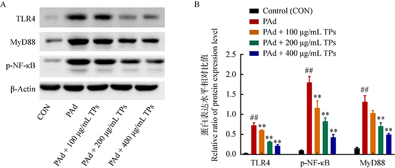
银耳多糖能够抑制棕榈酸诱导的Caco-2细胞促炎因子分泌,而炎症会导致肠道紧密连接蛋白受损(Li et al. 2019),因此,进一步检测经典的炎症通路TRL4/MyD88/NF-κB,探究银耳多糖是否通过影响TRL4/MyD88/NF-κB炎症信号通路来调控肠道屏障。PAd处理后TRL4、MyD88和p-NF-κB的表达量显著上调,TPs处理能有效抑制3种蛋白的表达,随着剂量升高抑制的效果越好(图6),说明TPs能够通过抑制TRL4/MyD88/NF-κB炎症信号通路下调促炎因子分泌。
图6 银耳多糖对TRL4/MyD88/NF-κB信号通路的影响 A:Western blot检测TLR4、MyD88、p-NF-κB的蛋白条带;B:TLR4、MyD88、p-NF-κB的相对表达量

Fig. 6 Effects of TPs on TRL4/MyD88/NF-κB signaling pathway. A: Protein bands of TLR4, MyD88, and p-NF-κB were detected by Western blot. B: Relative expression of TLR4, MyD88, and p-NF-κB.

Full size|PPT slide

3 讨论

肥胖症的流行在世界范围内不断蔓延,大大增加了其他相关慢性代谢疾病的全球负担,如2型糖尿病、非酒精性脂肪性肝病和心血管疾病等(James et al. 2018)。肥胖通常伴随着机体激素、炎症、脂质和葡萄糖水平紊乱的改变,这些代谢疾病都与肠道屏障息息相关(Zhang et al. 2024)。研究表明,多种来源的多糖能够维护肠道屏障进而干预代谢疾病。茯苓多糖可缓解由细胞焦亡驱动的肠道屏障破坏,从而减轻非酒精性脂肪性肝炎(Ye et al. 2022),黄芩多糖通过改善肠道屏障功能和调节肠道菌群改善葡聚糖硫酸钠诱导的溃疡性结肠炎(Cui et al. 2021),果胶多糖通过调节肠道菌群,增加肠道屏障功能和免疫增强活性,达到抑制肥胖、脂肪肝和炎症的效果(Lee et al. 2022)。
高脂饮食诱导的肥胖大鼠和小鼠肠道紧密连接蛋白Occludin、Claudin-1等蛋白的表达水平受到明显抑制(Suzuki & Hara 2010;Cremonini et al. 2019),作为食物中常见的饱和脂肪酸,摄入PAd的小鼠肠道紧密连接蛋白也明显减少或易位,从而影响肠道屏障功能(Xiao et al. 2017;Zhang et al. 2019;何亚伦等 2022)。细胞活力和完整性实验结果表明,暴露在PAd和TPs下不会对Caco-2细胞产生毒性作用,PAd能够损伤Caco-2细胞完整性,经TPs处理可以减轻PAd对细胞造成的损伤。
氧化还原平衡对维持肠道屏障至关重要,随着体内活性氧水平积累、内源性抗氧化物质消耗,以及氧化应激标志物水平增加,过量的活性氧导致氧化应激诱发炎症,分泌促炎因子(Ge et al. 2022;苏安祥等 2022)。经PAd处理后的Caco-2细胞抗氧化物SOD和CAT水平降低,脂质过氧化物MDA升高。由于硫氧还蛋白系统(TRX)、谷胱甘肽系统(GSH)以及核因子E2相关因子2 (NRF2)都参与调节细胞氧化还原稳态,目前尚不明确TPs是通过什么途径调控Caco-2细胞的氧化还原状态,可能是单一途径或者是多途径共同调控,因此,调控作用与剂量不成正比(Muri & Kopf 2020)。促炎因子IL-1β、TNF-α、IL-6分泌增加,说明PAd引发Caco-2细胞氧化应激反应并诱发了炎症,而TPs处理能有效缓解PAd由氧化应激诱发的炎症。促炎因子能够通过调控紧密连接蛋白的表达和组装,作用于紧密连接蛋白的功能。研究表明,Caco-2细胞与TNF-α和IFN-γ共同培养导致紧密连接蛋白ZO-1、Occludin、Claudin-1和Claudin-4重组,降低肠道屏障功能(Zolotarevsky et al. 2002)。Claudin-2是IL-22在肠上皮中作用的一个关键靶蛋白,IL-22能够诱导Claudin-2表达上调,进而提高肠道上皮屏障通透性(Ong et al. 2020)。经TPs处理能显著缓解PAd导致的紧密连接蛋白Claudin-1、Occludin和ZO-1表达量降低,说明PAd由氧化应激诱发的炎症影响紧密连接蛋白表达,而TPs能够通过提升抗氧化性能、抑制促炎因子分泌、提升紧密连接蛋白表达水平达到维护肠道屏障的作用。研究发现氢能够还原多余自由基调节氧化应激平衡,通过该方法调控TLR-4/MyD88/NF-κB信号通路最终保护肠道屏障(Li et al. 2024),NF-κB表达水平与肠上皮细胞凋亡息息相关,进而影响肠道屏障功能(Yao & Cadwell 2020;Yue et al. 2020)。因此,结合经典的TLR-4/MyD88/NF-κB信号通路,确认TPs能够通过抑制TRL4/MyD88/NF-κB炎症信号通路下调促炎因子分泌,进而保护肠道屏障。
课题组前期研究表明,TPs能够调控肠道菌群及菌群代谢物(Xu et al. 2021),而肠道菌群紊乱也会导致肠道屏障受损(雷棋怡等 2024),目前发现TPs可以干预PAd导致的肠道屏障受损,未来可进一步探究TPs改变的肠道菌群是否对肠道屏障产生影响,从而更全面地说明TPs对肠道屏障的作用。

4 结论

高剂量的TPs能够显著提升细胞完整性和抗氧化性,通过抑制TRL4/MyD88/NF-κB炎症信号通路抑制促炎因子分泌,提升紧密连接蛋白Claudin-1、Occludin和ZO-1表达,达到保护肠道屏障的作用。本文为揭示TPs维持肠道屏障的机制和扩展其应用范围提供了科学依据和思路。

作者贡献

董茜:实验、论文撰写;张仕林:实验、论文撰写;谢丽源:提供实验材料、验证;张谦:数据管理、软件作图、验证数据;舒雪琴:软件使用、作图;何晓兰:提供菌种和实验材料、修改论文;彭卫红:修改论文;许瀛引:论文构思、修改论文。

利益冲突

作者声明,该研究不存在任何潜在利益冲突的商业或财务关系。

参考文献

[1]
Campbell EL, Colgan SP, 2019. Control and dysregulation of redox signalling in the gastrointestinal tract. Nature Reviews Gastroenterology & Hepatology, 16(2): 106-120
[2]
Cremonini E, Daveri E, Mastaloudis A, Adamo AM, Mills D, Kalanetra K, Hester SN, Wood SM, Fraga CG, Oteiza PI, 2019. Anthocyanins protect the gastrointestinal tract from high fat diet-induced alterations in redox signaling, barrier integrity and dysbiosis. Redox Biology, 26: 101269
[3]
Cui L, Guan X, Ding W, Luo Y, Wang W, Bu W, Song J, Tan X, Sun E, Ning Q, Liu G, Jia X, Feng L, 2021. Scutellaria baicalensis Georgi polysaccharide ameliorates DSS-induced ulcerative colitis by improving intestinal barrier function and modulating gut microbiota. International Journal of Biological Macromolecules, 1(166): 1035-1045
[4]
Ge L, Qi J, Shao B, Ruan Z, Ren Y, Sui S, Wu X, Sun X, Liu S, Li S, Xu C, Song W, 2022. Microbial hydrogen economy alleviates colitis by reprogramming colonocyte metabolism and reinforcing intestinal barrier. Gut Microbes, 14(1): 2013764
[5]
Ge X, Huang W, Xu X, Lei P, Sun D, Xu H, Li S, 2020. Production, structure, and bioactivity of polysaccharide isolated from Tremella fuciformis XY. International Journal of Biological Macromolecules, 148: 173-181
[6]
Genser LD, Aguanno HAS, Dong L, Trystram L, Assmann K, Salem JE, Vaillant JC, Oppert JM, Laugerette F, Michalski MC, Wind P, Rousset M, Brot-Laroche E, Leturque A, Clément K, Thenet S, Poitou C, 2018. Increased jejunal permeability in human obesity is revealed by a lipid challenge and is linked to inflammation and type 2 diabetes. Journal of Pathology, 246(2): 217-230
[7]
Ghezzal S, Postal BG, Quevrain E, Brot L, Seksik P, Leturque A, Thenet S, Carrière V, 2020. Palmitic acid damages gut epithelium integrity and initiates inflammatory cytokine production. Biochimica et Biophysica Acta-Molecular and Cell Biology of Lipids, 1865(2): 158530
[8]
He YL, Zeng LR, Liu X, Zhang L, Wang Q, 2022. Effects of high doses of tannin on intestinal barrier and intestinal flora in mice. Biotechnology Bulletin, 38(4): 278-287 (in Chinese)
[9]
James SL, Abate D, Abate KH, et al. (more than 20 authors), 2018. Global, regional, and national incidence, prevalence, and years lived with disability for 354 diseases and injuries for 195 countries and territories, 1990-2017: a systematic analysis for the Global Burden of Disease Study 2017. Lancet, 392(10159): 1789-1858
[10]
Kawai T, Autieri MV, Scalia R, 2021. Adipose tissue inflammation and metabolic dysfunction in obesity. American Journal of Physiology-Cell Physiology, 320(3): C375-C391
[11]
Lee HB, Kim YS, Park HY, 2022. Pectic polysaccharides: targeting gut microbiota in obesity and intestinal health. Carbohydrate Polymers, 1(287): 119363
[12]
Lei QY, Xu Y, Li PF, 2024. Effect and mechanism of Bacteroides fragilis type Ⅵ secretion system on intestinal barrier. Biotechnology Bulletin, 40(3): 286-295 (in Chinese)
[13]
Lerner A, Matthias T, 2015. Changes in intestinal tight junction permeability associated with industrial food additives explain the rising incidence of autoimmune disease. Autoimmunity Reviews, 14(6): 479-489
The incidence of autoimmune diseases is increasing along with the expansion of industrial food processing and food additive consumption. The intestinal epithelial barrier, with its intercellular tight junction, controls the equilibrium between tolerance and immunity to non-self-antigens. As a result, particular attention is being placed on the role of tight junction dysfunction in the pathogenesis of AD. Tight junction leakage is enhanced by many luminal components, commonly used industrial food additives being some of them. Glucose, salt, emulsifiers, organic solvents, gluten, microbial transglutaminase, and nanoparticles are extensively and increasingly used by the food industry, claim the manufacturers, to improve the qualities of food. However, all of the aforementioned additives increase intestinal permeability by breaching the integrity of tight junction paracellular transfer. In fact, tight junction dysfunction is common in multiple autoimmune diseases and the central part played by the tight junction in autoimmune diseases pathogenesis is extensively described. It is hypothesized that commonly used industrial food additives abrogate human epithelial barrier function, thus, increasing intestinal permeability through the opened tight junction, resulting in entry of foreign immunogenic antigens and activation of the autoimmune cascade. Future research on food additives exposure-intestinal permeability-autoimmunity interplay will enhance our knowledge of the common mechanisms associated with autoimmune progression.Copyright © 2015. Published by Elsevier B.V.
[14]
Li J, Huang G, Wang J, Wang S, Yu Y, 2024. Hydrogen regulates ulcerative colitis by affecting the intestinal redox environment. Journal of Inflammation Research, 17: 933-945
The redox balance in the intestine plays an important role in maintaining intestinal homeostasis, and it is closely related to the intestinal mucosal barrier, intestinal inflammation, and the gut microbiota. Current research on the treatment of ulcerative colitis has focused on immune disorders, excessive inflammation, and oxidative stress. However, an imbalance in intestinal redox reaction plays a particularly critical role. Hydrogen is produced by some anaerobic bacteria via hydrogenases in the intestine. Increasing evidence suggests that hydrogen, as an inert gas, is crucial for immunity, inflammation, and oxidative stress and plays a protective role in ulcerative colitis. Hydrogen maintains the redox state balance in the intestine in ulcerative colitis and reduces damage to intestinal epithelial cells by exerting its selective antioxidant ability. Hydrogen also regulates the intestinal flora, reduces the harmful effects of bacteria on the intestinal epithelial barrier, promotes the restoration of normal anaerobic bacteria in the intestines, and ultimately improves the integrity of the intestinal epithelial barrier. The present review focuses on the therapeutic mechanisms of hydrogen-targeting ulcerative colitis.© 2024 Li et al.
[15]
Li Y, Tian X, Li S, Chang L, Sun P, Lu Y, Yu X, Chen S, Wu Z, Xu Z, Kang W, 2019. Total polysaccharides of adlay bran (Coix lachryma-jobi L.) improve TNF-α induced epithelial barrier dysfunction in Caco-2 cells via inhibition of the inflammatory response. Food Function, 10(5): 2906-2913
[16]
Miao S, Mao X, Pei R, Miao S, Xiang C, Lv Y, Yang X, Sun J, Jia S, Liu Y, 2013. Antitumor activity of polysaccharides from Lepista sordida against laryngocarcinoma in vitro and in vivo. International Journal of Biological Macromolecules, 60: 235-240
[17]
Miner-Williams WM, Moughan PJ, 2016. Intestinal barrier dysfunction: implications for chronic inflammatory conditions of the bowel. Nutrition Research Reviews, 29(1): 40-59
The intestinal epithelium of adult humans acts as a differentially permeable barrier that separates the potentially harmful contents of the lumen from the underlying tissues. Any dysfunction of this boundary layer that disturbs the homeostatic equilibrium between the internal and external environments may initiate and sustain a biochemical cascade that results in inflammation of the intestine. Key to such dysfunction are genetic, microbial and other environmental factors that, singularly or in combination, result in chronic inflammation that is symptomatic of inflammatory bowel disease (IBD). The aim of the present review is to assess the scientific evidence to support the hypothesis that defective transepithelial transport mechanisms and the heightened absorption of intact antigenic proinflammatory oligopeptides are important contributing factors in the pathogenesis of IBD.
[18]
Mineroff J, Jagdeo J, 2023. The potential cutaneous benefits of Tremella fuciformis. Archives of Dermatological Research, 315(7): 1883-1886
Tremella fuciformis, also known as snow mushroom, is an edible mushroom that has historically been popular in herbal and Asian medicine and cuisine. The main polysaccharide ingredients have been extracted and used as treatment in a variety of conditions, demonstrating positive effects in a range of biological functions including those involved in antioxidation, antitumor, antidiabetic, immunomodulatory, and neuroprotective pathways. Studies have demonstrated the role this extract may play in skin antiaging, photoprotection, wound healing, and barrier protection. Most studies have been limited to in vitro and in vivo animal models. Future clinical research is needed to further understand the role of T. fuciformis in dermatology. This review will discuss the existing research findings and potential future applications for T. fuciformis as a treatment in skin conditions.© 2023. The Author(s), under exclusive licence to Springer-Verlag GmbH Germany, part of Springer Nature.
[19]
Muri J, Kopf M, 2020. Redox regulation of immunometabolism. Nature Reviews Immunology, 21: 363-381
[20]
Ohata A, Usami M, Miyoshi M, 2005. Short-chain fatty acids alter tight junction permeability in intestinal monolayer cells via lipoxygenase activation. Nutrition, 21(7-8): 838-847
[21]
Ong M, Yeruva S, Sailer A, Nilsen SP, Turner JR, 2020. Differential regulation of claudin-2 and claudin-15 expression in children and adults with malabsorptive disease. Laboratory Investigation, 100(3): 483-490
Intestinal Na-nutrient cotransport depends on claudin-2 and claudin-15 mediated Na recycling. Expression of these proteins is coordinately regulated during postnatal development. While expression of claudin-2 and claudin-15 has been studied in inflammatory bowel disease (IBD) and celiac disease (CD), it has not been assessed in other malabsorptive diseases, and no reports have compared expression in children and adults. We used quantitative immunofluorescence microscopy to assess claudin-2 and claudin-15 expression in duodenal biopsies from children and adults with malabsorptive disease and healthy controls. Consistent with previous work in rodents, claudin-2 expression in healthy children was markedly greater, and claudin-15 expression was less, than that in adults. Claudin-2 expression was increased in adults with CD and downregulated in children with graft-versus-host disease (GVHD). In contrast, claudin-15 expression was reduced in adults with GVHD and common variable immunodeficiency (CVID). These data show that one of the two Na/water pore-forming claudins is upregulated in CD and downregulated in GVHD and CVID. The specific claudin whose expression changes, however, reflects the age of the patient (child or adult). We conclude that contributions of claudin-2 and claudin-15 to pathophysiology of and responses to diarrhea in children and adults with GVHD and CVID differ from those in CD and IBD.
[22]
Su AX, Hu Y, Hu QH, Xu H, Liu JH, Xie MH, Pei F, Yang WJ, 2022. The inhibitory effect of Flammulina velutipes proteoglycan on inflammation induced by lipopolysaccharide in Caco-2/RAW264.7 cell co-culture model. Food Science, 43(3): 146-151 (in Chinese)
[23]
Suzuki T, 2020. Regulation of the intestinal barrier by nutrients: the role of tight junctions. Journal of Animal Science, 91(1): e13357
[24]
Suzuki T, Hara H, 2010. Dietary fat and bile juice, but not obesity, are responsible for the increase in small intestinal permeability induced through the suppression of tight junction protein expression in LETO and OLETF rats. Nutrition & Metabolism, 7: 19
[25]
Tilg H, Zmora N, Adolph TE, Elinav E, 2020. The intestinal microbiota fuelling metabolic inflammation. Nature Reviews Immunology, 20(1): 40-54
Low-grade inflammation is the hallmark of metabolic disorders such as obesity, type 2 diabetes and nonalcoholic fatty liver disease. Emerging evidence indicates that these disorders are characterized by alterations in the intestinal microbiota composition and its metabolites, which translocate from the gut across a disrupted intestinal barrier to affect various metabolic organs, such as the liver and adipose tissue, thereby contributing to metabolic inflammation. Here, we discuss some of the recently identified mechanisms that showcase the role of the intestinal microbiota and barrier dysfunction in metabolic inflammation. We propose a concept by which the gut microbiota fuels metabolic inflammation and dysregulation.
[26]
Xiao YT, Yan WH, Cao Y, Yan JK, Cai W, 2017. P 38 MAPK pharmacological inhibitor SB203580 alleviates total parenteral nutrition-induced loss of intestinal barrier function but promotes hepatocyte lipoapoptosis. Cellular Physiology and Biochemistry, 41(2): 623-634
[27]
Xu Y, Xie L, Zhang Z, Zhang W, Tang J, He X, Zhou J, Peng W, 2021. Tremella fuciformis polysaccharides inhibited colonic inflammation in dextran sulfate sodium-treated mice via Foxp3+ T cells, gut microbiota, and bacterial metabolites. Frontiers in Immunology, 12: 648162
[28]
Yang D, Liu Y, Zhang L, 2019. Tremella polysaccharide: the molecular mechanisms of its drug action. Progress in Molecular Biology and Translational Science, 163: 383-421
Tremella fuciformis is an edible medicinal mushroom well known as "Yiner" or "Baimuer" in China and has been used as a Chinese herb for many years. T. fuciformis polysaccharide (TFPS) has been identified as a major bioactive component. Different experimental conditions can obtain different TFPS fractions, which makes TFPS a mixture of different polysaccharides with the molecular weight ranging from 5.82×10Da to 3.74×10Da. The monosaccharides detected in TFPS include mannose, xylose, fucose, glucuronic acid, glucose, and galactose. One characterized TFPS chemical structure consists of a linear (1→3)-linked α-d-mannose backbone with highly branched β-d-xylose, α-d-fucose and β-d-glucuronic acid as the side chains. TFPS shows multiple physiological and healthy promoting effects including immunomodulation, antitumor, anti-oxidation, anti-aging, hypoglycemic, hypolipidemic, neuroprotection, and other effects. As a result, "Tremella Polysaccharide Enteric-coated Capsules" was approved by Chinese Food and Drug Administration (SFDA) in 2002 for treating cancer patients with leukopenia induced by chemotherapy and radiotherapy. It is also used as adjuvant drug for treating chronic persistent hepatitis and chronic active hepatitis. In this chapter, 113 independent studies involving in biochemical, pharmacological, and clinical studies of TFPS during the past 46 years (1972-2018) on the base of PubMed, CNKI (China National Knowledge Infrastructure) and Wanfang database search are summarized. TFPS shows efficacy for all types of human diseases in the reported clinical studies. The structure, molecular mechanisms of the immunomodulation, antitumor, anti-oxidation, anti-aging, hypoglycemic, hypolipidemic, preclinical and clinical efficacy are discussed to provide a general picture of TFPS as a clinically used drug.© 2019 Elsevier Inc. All rights reserved.
[29]
Yao X, Cadwell K, 2020. Tumor necrosis factor-α-induced apoptosis in the intestinal epithelium due to chronic nuclear factor kappa B signaling is mediated by receptor interacting serine/threonine kinase 1. Cellular and Molecular Gastroenterology and Hepatology, 9(2): 337-338
[30]
Ye H, Ma S, Qiu Z, Huang S, Deng G, Li Y, Xu S, Yang M, Shi H, Wu C, Li M, Zhang J, Zhang F, Qin M, Huang H, Zeng Z, Wang M, Chen Y, Lin H, Gao Z, Cai M, Song Y, Gong S, Gao L, 2022. Poria cocos polysaccharides rescue pyroptosis-driven gut vascular barrier disruption in order to alleviates non-alcoholic steatohepatitis. Journal of Ethnopharmacology, 5(296): 115457
[31]
Yue B, Ren J, Yu Z, Luo X, Ren Y, Zhang J, Mani S, Wang Z, Dou W, 2020. Pinocembrin alleviates ulcerative colitis in mice via regulating gut microbiota, suppressing TLR4/MD2/NF-κB pathway and promoting intestinal barrier. Bioscience Reports, 40(7): BSR20200986
[32]
Zeisel MB, Dhawan P, Baumert TF, 2019. Tight junction proteins in gastrointestinal and liver disease. Gut: Journal of the British Society of Gastroenterology, 68(3): 547-561
[33]
Zhang J, Huang Y, Li H, Xu P, Liu Q, Sun Y, Zhang Z, Wu T, Tang Q, Jia Q, Xia Y, Xu Y, Jing X, Li J, Mo L, Xie W, Qu A, He J, Li Y, 2024. B3galt 5 functions as a PXR target gene and regulates obesity and insulin resistance by maintaining intestinal integrity. Nature Communication, 15(1): 5919
[34]
Zhang P, Yu Y, Qin Y, Zhou Y, Tang R, Wang Q, Li X, Wang H, Weston-Green K, Huang XF, Zheng K, 2019. Alterations to the microbiota-colon-brain axis in high-fat-diet-induced obese mice compared to diet-resistant mice. Journal of Nutritional Biochemistry, 65: 54-65
Obesity is underpinned by both genetic and environmental factors, including a high-saturated-fat diet. Some mice develop diet-induced obesity (DIO), but others remain diet resistant (DR) despite intake of the same high-saturated-fat diet, a phenomenon that mimics characteristics of the human obese phenotype. Microbiota-colon-brain axis regulation is important for energy metabolism and cognition. Using DIO and DR mouse models, this study aimed to examine gut microbiota, colonic inflammation and cognitive function to elucidate the role of microbiota-gut-brain regulation in DIO. C57Bl6/J mice fed a chronic saturated-palmitic-acid diet for 22 weeks showed significant body weight gain differences, with the top one third gaining 48% heavier body weight than the lower one third. There was significant reduction in gut microbiota richness and diversity in DIO mice but not in DR mice. At the phylum level, DIO mice had increased abundance of Firmicutes and Antinobacteria, and decreased abundance of Bacterioides and Proteobacteria in gut microbiota. DIO mice exhibited reduced tight junction proteins, increased plasma endotoxin lipopolysaccharide (LPS) and increased inflammation in the colon and liver. Recognition memory and spatial memory were impaired in DIO mice, associated with decreased Bacteroidetes. Further examination showed that hippocampal brain-derived neurotrophic factor was significantly decreased in DIO mice (vs. DR). Conversely, DR mice showed no changes in the above parameters measured. Therefore, gut microbiota, colon inflammation and circulating LPS may play a major role in the development of the obese phenotype and cognitive decline associated with a chronic high-saturated-palmitic-acid diet.Copyright © 2018. Published by Elsevier Inc.
[35]
Zolotarevsky Y, Hecht G, Koutsouris A, Gonzalez DE, Quan C, Tom J, Mrsny RJ, Turner JR, 2002. A membrane-permeant peptide that inhibits MLC kinase restores barrier function in vitro models of intestinal disease. Gastroenterology, 123(1): 163-172
Maintenance of the mucosal barrier is a critical function of intestinal epithelia. Myosin regulatory light chain (MLC) phosphorylation is a common intermediate in the pathophysiologic regulation of this barrier. The aim of this study was to determine whether a membrane permeant inhibitor of MLC kinase (PIK) could inhibit intracellular MLC kinase and regulate paracellular permeability.Recombinant MLC and Caco-2 MLC kinase were used for kinase assays. T84 and Caco-2 monolayers were treated with enteropathogenic Escherichia coli (EPEC) or tumor necrosis factor (TNF)-alpha and interferon (IFN)-gamma to induce barrier dysfunction.PIK inhibited MLC kinase in vitro and was able to cross cell membranes and concentrate at the perijunctional actomyosin ring. Consistent with these properties, apical addition of PIK reduced intracellular MLC phosphorylation by 22% +/- 2%, increased transepithelial resistance (TER) by 50% +/- 1%, and decreased paracellular mannitol flux rates by 5.2 +/- 0.2-fold. EPEC infection induced TER decreases of 37% +/- 6% that were limited to 16% +/- 5% by PIK. TNF-alpha and IFN-gamma induced TER decreases of 22% +/- 3% that were associated with a 172% +/- 1% increase in MLC phosphorylation. Subsequent PIK addition caused MLC phosphorylation to decrease by 25% +/- 4% while TER increased to 97% +/- 6% of control.PIK can prevent TER defects induced by EPEC and reverse MLC phosphorylation increases and TER decreases induced by TNF-alpha and IFN-gamma. The data also suggest that TNF-alpha and IFN-gamma regulate TER, at least in part, via the perijunctional cytoskeleton. Thus, PIK may be the prototype for a new class of targeted therapeutic agents that can restore barrier function in intestinal disease states.
[36]
何亚伦, 曾丽荣, 刘雄, 张铃, 王琼, 2022. 高剂量单宁酸对小鼠肠道屏障和肠道菌群的影响. 生物技术通报, 38(4): 278-287
以正常饮食小鼠和高脂饮食诱导肥胖模型小鼠为对象,研究高剂量单宁酸对小鼠肠道屏障及肠道菌群的影响。通过H&#x00026;E染色、RT-qPCR、16S rRNA测序等方法进行检测分析。研究发现高剂量单宁酸(400 mg/kg BW)灌胃可使小鼠的体重和进食量降低,增加小鼠体内各肠段内容物的含量,其中结肠内容物的含量显著增加。并且高剂量单宁酸干预后可造成肠道功能损伤及肠道屏障破坏,如杯状细胞数量和隐窝长度减少,及肠道紧密连接蛋白(ZO-1,Occludin及Claudin)表达降低。此外,口服高剂量单宁酸会导致小鼠结肠肠道菌群的多样性发生变化,并增加了SCFA产生菌Alistipes,Ruminococcus和 Blautia以及肥胖负相关菌群Alistipes和Oscillibacter的含量。结果表明,高剂量单宁酸对肠黏膜屏障的破坏影响了小鼠对食物的消化吸收,这可能是高剂量单宁酸干预后小鼠体重快速下降的主要潜在原因。另一方面,高剂量单宁酸所造成的肠道菌群变化也会对小鼠的体重产生影响。
[37]
雷棋怡, 徐杨, 李鹏飞, 2024. 脆弱拟杆菌六型分泌系统对肠道屏障的影响及机制. 生物技术通报, 40(3): 286-295
【目的】 探讨脆弱拟杆菌(Bacteroides fragilis)中六型蛋白分泌系统(T6SS)对肠道屏障的影响及作用机制。【方法】 采用自杀载体构建B. fragilis T6SS缺陷株。建立葡聚糖硫酸钠(DSS)诱导的肠炎小鼠模型,并分别补充PBS,B. fragilis WT和B. fragilis ΔT6SS,随后比较三组小鼠疾病特征、肠道屏障完整性的差异。利用荧光定量PCR和免疫组化检测小鼠紧密连接蛋白的表达。采用非靶向代谢组学比较各组小鼠肠道差异代谢物。【结果】 T6SS缺失不影响B. fragilis的生物活性。与PBS对照组相比,B. fragilis WT明显改善小鼠的体重丢失、结肠长度等疾病指标,表现出对DSS诱导的肠炎的保护性,而B. fragilis ΔT6SS随着T6SS的敲除丧失了对DSS诱导的肠炎的保护性。小鼠血清中异硫氰酸荧光素葡聚糖含量和肠道病理切片均表明B. fragilis T6SS能改善小鼠肠道屏障的完整性。荧光定量PCR和免疫组化均表明B. fragilis T6SS影响肠道紧密连接蛋白的表达。非靶向代谢组学分析表明,与B. fragilis ΔT6SS组相比,B. fragilis WT组显著上调96个肠道差异代谢产物,其中多个代谢产物富集到胆碱能突触代谢和甘油磷脂代谢相关通路。【结论】 拟杆菌T6SS改变肠道代谢组并提高肠道细胞紧密连接蛋白的表达,改善肠道屏障的通透性,参与到拟杆菌对肠道屏障的保护性。
[38]
苏安祥, 胡烨, 胡秋辉, 徐辉, 刘建辉, 谢旻皓, 裴斐, 杨文建, 2022. 金针菇蛋白聚糖对脂多糖诱导的Caco-2/RAW264.7细胞共培养模型炎症的抑制作用. 食品科学, 43(3): 146-151

基金

国家自然科学基金(32402653)
四川省自然科学基金(2023NSFSC1249)
四川省财政自主创新专项(2022ZZCX096)
PDF(782 KB)

73

Accesses

0

Citation

Detail

段落导航
相关文章

/