PDF(827 KB)

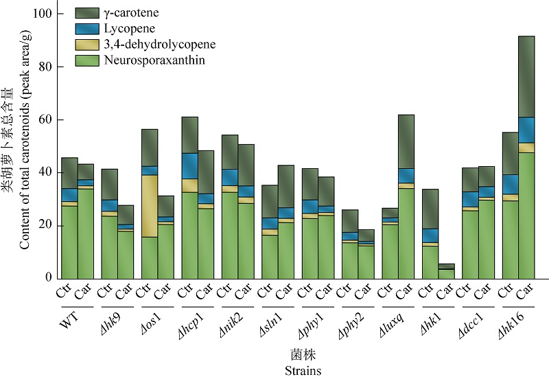
Mechanism of histidine kinases in responses to carvacrol stress in Neurospora crassa
CHEN Pengxu, LAN Ziyi, XI Juan, CHEN Yingying, ZHENG Weifa, ZHAO Yanxia
Mycosystema ›› 2025, Vol. 44 ›› Issue (4) : 240268.
PDF(827 KB)
PDF(827 KB)
Mechanism of histidine kinases in responses to carvacrol stress in Neurospora crassa
 ({{custom_author.role_en}})
,
{{javascript:window.custom_author_en_index++;}}
({{custom_author.role_en}})
,
{{javascript:window.custom_author_en_index++;}}
| {{custom_ref.label}} |
{{custom_citation.content}}
{{custom_citation.annotation}}
|


/
| 〈 |
|
〉 |