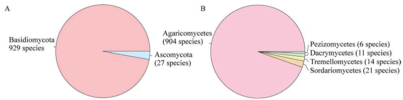
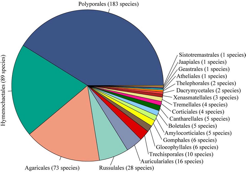
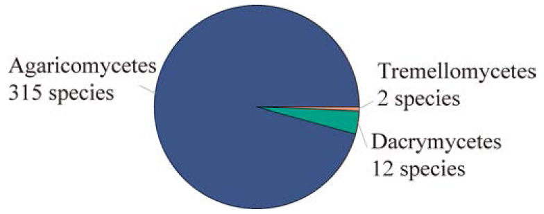
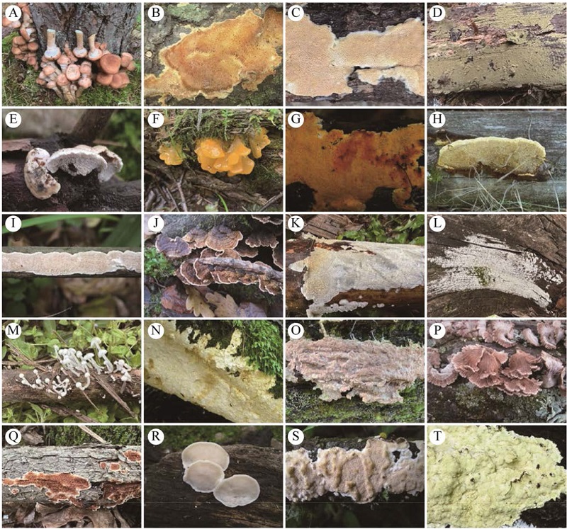
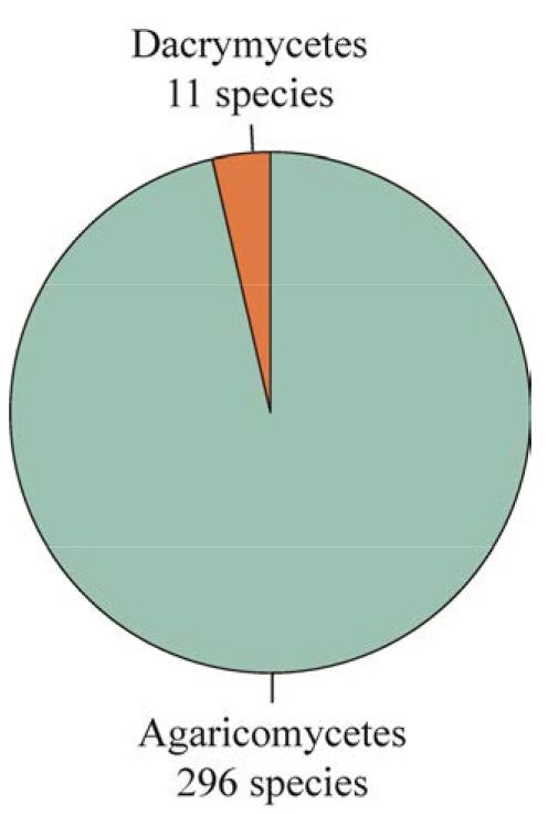
Guangxi Autonomous Region is characterized by complex topography and a subtropical climate in southern China, which harbors rich macrofungal diversity. This study compiled the species diversity of wood-decaying fungi in Guangxi by integrating datasets from four institutes, particularly the identification of more than 6 000 specimens collected during 2023-2025. A total of 956 species is recorded, representing 2 phyla, 5 classes, 21 orders, 97 families, and 337 genera, and the phylum Basidiomycota (Agaricomycetes) dominates the assemblage. Among these, Polyporales, Hymenochaetales, Agaricales, and Russulales are dominant classes, with the sum of species accounting for 85.25% of the total known species. In total, 11 dominant families and 12 dominant genera are recognized, representing 55.96% and 20.61% of the total known species, respectively. In addition, 70 species (7.32% of the total known species) were described on the basis of holotypes or paratypes sampled from Guangxi. This study provides a comprehensive overview of the species composition and diversity patterns of wood-decaying fungi in Guangxi.
Wood-decaying fungi as a group of macrofungi have significant economic values and ecological functions. Zhangguangcai Mountain, located in borderland of Heilongjiang and Jilin provinces, are one of the main mountain ranges and forest areas in Northeast China, providing an important ecological niche for the growth of wood decay fungi. The species diversity of wood-decaying fungi in the mountains was poorly known, therefore a systematic investigation was conducted. Based on the identification of related specimens from the mountains preserved in major domestic herbaria, a total of 445 species is identified; among them 345 species are reported for the first time reported in the mountains. The known 445 species belong to three classes, 20 orders, 82 families, and 239 genera, within Basidiomycota. The dominant orders are Polyporales, Hymenochaetales, and Agaricales, and the sum of species accounts for 77.53% of all wood-decaying fungal species recorded in the mountain range. The dominant families are Polyporaceae, Hymenochaetaceae, Schizoporaceae, Steccherinaceae, and Meruliaceae, and the sum of species accounts for 32.13% of the total known species in the areas. The dominant genera are Xylodon, Trametes, Skeletocutis, Steccherinum, Cyanosporus, Lyomyces, Phellinus, Antrodiella, Peniophorella, and Trechispora, with the sum of species constituting 16.85% of the total known species of wood-decaying fungi in the areas.
Pinus massoniana has wide distribution in the subtropical regions of China with important economic values. Although several studies have previously carried out on the fungal diversity associated with P. massoniana, a comprehensive systematic assessment is still lacking. The present study compiles the wood-decomposing fungi on P. massoniana based on identification of collected samples, published literatures and herbarium specimen data. A total of 329 species, belonging to 3 classes, 16 orders, 72 families, and 169 genera within Basidiomycota, is recorded, of which more than 70% of these species are newly recorded on wood of P. massoniana. The dominant orders are Polyporales, Hymenochaetales, and Agaricales, with the sum of species accounting for 78.72% of the total species. The dominant families include Polyporaceae, Schizoporaceae, Incrustoporiaceae, Meruliaceae, Hymenochaetaceae, Phanerochaetaceae, Irpicaceae, Postiaceae, Dacrymycetaceae, and Fomitopsidaceae, with the sum of species representing 48.33% of the total species. The dominant genera are Skeletocutis, Xylodon, Peniophorella, Cyanosporus, Dacrymyces, Hyphoderma, Trametes, and Trechispora, accounting for 19.15% of the total species. This study reveals the species composition and community structure of wood-decomposing fungi associated with P. massoniana, enriching the knowledge of fungal biodiversity in China and providing reference material for forest protection and utilization.
Pinus tabulaeformis is a unique evergreen and one of the most important forest tree species in China. Although some previous reports are involved with certain species of wood-inhabiting fungi on the tree, these seem fragmentary and unsystematic. Based on identification of our collected specimens, related literature and the data of herbaria, systematic sort-out and compilation of wood decay fungi on P. tabulaeformis is carried out. In total, 305 species of belonging to 144 genera, 56 families, and 14 orders are recorded. 74% of these species are reported for the first time on P. tabulaeformis; especially, most of the corticioid species are reported for the first time on the tree. The dominant orders are Polyporales, Hymenochaetales, and Agaricales, with the sum of species accounting for 78.72% of the total number of wood-decaying species. These three orders included 36 families, 107 genera and 235 species, which accounted for 64.29%, 74.31% and 77.05% of the total family, genera and species respectively. The dominant families are Polyporaceae, Phanerochaetaceae, Schizoporaceae, Hydnodontaceae, Hymenochaetaceae, Incrustoporiaceae, Irpicaceae and Omphalotaceae, and the sum of genera of these eight families accounted for 30.56% of the total number of wood-decaying genera, while the sum of species of these eight families accounted for 39.67% of the total species. In addition, there are ten dominant genera, with sum of species accounting for 25.57% of the total species. This is the first catalogue of wood-decaying fungi on P. tabulaeformis in China, providing basic data for research and utilization of the natural resource.
Pinus yunnanensis is one of the most important coniferous tree species in southwestern China, providing essential habitats for fungi. However, studies on species diversity of wood-decaying fungi associated with P. yunnanensis are still lacking. In this study, an integrated assessment of species composition and community characteristics of wood-decaying fungi on P. yunnanensis was conducted based on field specimen collection and identification, herbarium records, and reliable literature data. A total of 307 species of wood-decaying fungi was recorded, belonging to 2 classes, 14 orders, 64 families, and 143 genera within Basidiomycota. Polyporales and Hymenochaetales were the dominant orders, with the sum of species accounting for 67.43% of the total wood-decaying species. Nine dominant families were recognized, namely Schizoporaceae, Phanerochaetaceae, Fomitopsidaceae, Polyporaceae, Postiaceae, Hymenochaetaceae, Incrustoporiaceae, Dacrymycetaceae, and Irpicaceae, with the sum of species accounting for 43% of the total wood-decaying species. Seven dominant genera were identified, namely Xylodon, Skeletocutis, Peniophorella, Antrodia, Botryobasidium, Dacrymyces, and Phanerochaete, accounting for 20.20% of the total species. This study systematically reveals wood-decaying fungal diversity associated with Pinus yunnanensis in China, providing a reliable scientific basis for further exploration and utilization of the fungal resources.
Macrofungi are the key biological components in forest ecosystem, with some species having important economic values used as food, medicine and industry material. As the largest natural forest area in Beijing, Labagoumen National Forest Park has a typical forest ecosystem of the Yanshan Mountains, and its biodiversity is particularly abundant. In the present study, species diversity and funga composition of macrofungi occurring in the forest park were systematically investigated. In total, 316 species belonging to 21 orders, 72 families and 152 genera were recorded in this area, including 28 edible species, 14 medicinal species, 23 species concurrently edible and medicinal, and 42 toxic species. The funga conformed to the North Temperate distribution characteristics. Additionally, a new species, Singerocybe beijingensis, was found.
Database/Dataset Profile
| Title | A dataset on the species diversity and funga composition of macrofungi in Labagoumen National Forest Park, Beijing |
|---|---|
| Data author (s) | SHANG Zhirui, WU Yingda, YUAN Mingxuan, LI Zhongfeng, JI Yingyi, MA Jinxin, JIN Can, WANG Hao, SI Jing, LI Haijiao |
| Data corresponding author | SI Jing (jingsi1788@126.com); LI Haijiao (lihaijiao715@126.com) |
| Database/Dataset composition | The dataset consists of the results of a five-year continuous survey of macrofungi in Labagoumen National Forest Park, Beijing, as well as the analysis of the funga composition. |
| Time range (data generation time) | 2021−2025 |
| Geographical scope | Geographical scope: 39°23′N−41°05′N, 115°20′E−117°32′E |
| File size | 1 000 KB |
| Data volume | 21 Orders, 72 families, 152 genera and 316 species |
| Data format | .doc |
| Data link | Website ( |
Bamboo is an important economic plant in Yunnan and exhibits high species diversity. The genus Phyllachora is rich in species diversity and widely distributed worldwide. Phyllachora species are common on bamboos, however, the diversity of the species associated with bamboos remains poorly understood. Based on multi-locus phylogenetic analyses of ITS, LSU, and tef1-α together with morphological characteristics, two new species on bamboo, Phyllachora yushaniae-falcatiauritae on Yushania falcatiaurita and P. yushaniae-polytrichae on Y. polytricha are described and illustrated.
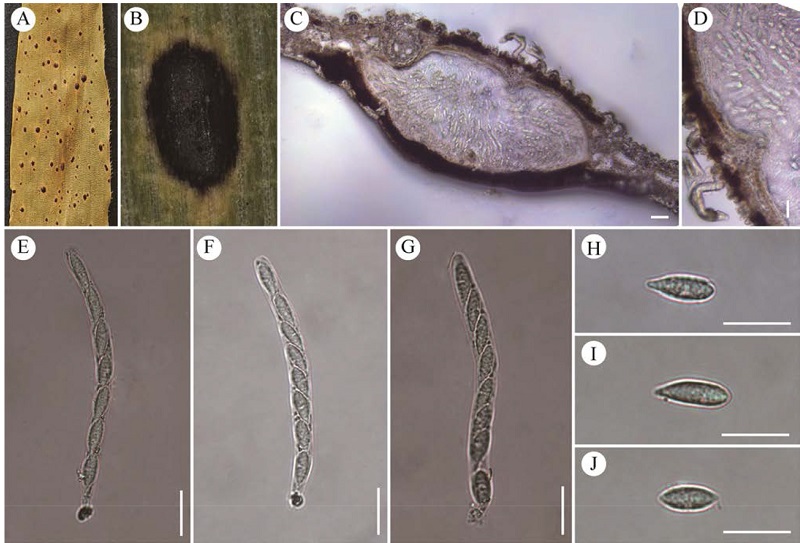
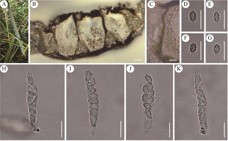
Based on multi-locus phylogenetic analyses of ITS and LSU together with morphological characteristics, a new species of Phyllachora on grass in the subfamily Eragrostoideae of the family Poaceae, P. eragrostidis-nigrae on Eragrostis nigra is described and illustrated. P. eragrostidis-nigrae is characterized by its ellipsoidal to ovoid ascospores (10-14 × 5-8 μm). In addition, Chloris anomala is reported as a new host for P. chloridis-virgatae.
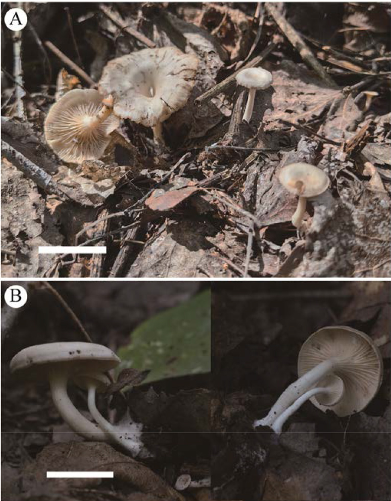
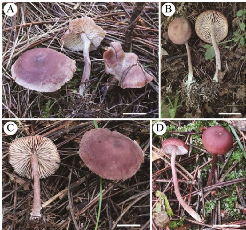
Xerophorus purpureus sp. nov., the first species of the genus Xerophorus recorded in China, is described and illustrated based on collections from Chifeng City, Inner Mongolia, Northeast China. The new species is characterized by its distinctive purple basidiomata, amygdaliform to ellipsoid basidiospores, and the presence of clamp connections. Phylogenetic analyses based on a combined four-locus dataset (ITS-SSU-LSU-rpb2) strongly supported X. purpureus as a distinct lineage.
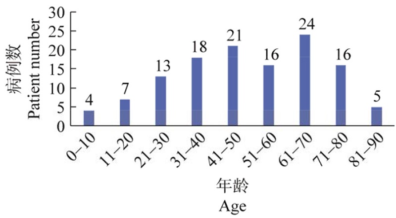
Vasculitis is a group of disorders in which inflammation injures the vessel wall and leads to structural changes. It can be triggered by immune-mediated inflammation, drugs, or infections. Among infectious causes, bacteria are most common, while the link between fungal infections and vasculitis has received less attention. Fungal-related vascular injury can be broadly categorized into two settings: opportunistic fungal infection occurring in primary immune-mediated vasculitis, and secondary vascular destruction caused by direct fungal angioinvasion of the vessel wall. According to PRISMA principles, PubMed and major Chinese databases from inception to December 31, 2024 were searched, and the case reports were included. Demographics, evidence supporting the vasculitis diagnosis, mycological evidence, treatment, and outcomes were extracted, and the cases were classified into primary immune-mediated vasculitis and secondary fungal vasculitis. A standardized framework was applied to grade the certainty of vasculitis diagnosis, the verification of mycological evidence, and the anatomical or temporal relevance between fungal infection and vascular lesions, generating an overall evidence level. Descriptive analyses were performed. Among cases with complete data on immunosuppression and mortality, mortality was compared using the chi-square test with Fisher’s exact test as a sensitivity check, and the risk ratio with 95% confidence intervals was calculated. In total, 106 reports involving 124 patients were included, covering 34 genera and 57 fungal species. The most frequently reported genera were Aspergillus, Candida, Coccidioides, Cryptococcus, Pneumocystis, and Histoplasma. Twenty vasculitis or vascular involvement phenotypes were identified, including most common ANCA-associated vasculitis, intracranial arteritis or arterial thrombosis, aneurysm, aortitis, thrombophlebitis, and transplant arteritis or transplant aneurysm. Mechanism-based stratification identified 40 cases of primary immune-mediated vasculitis with concomitant fungal infection and 84 cases of fungal infection-related secondary vascular lesions. Age ranged from 3 to 87 years, with the highest frequency in patients aged 61 to 70 years. Mortality analysis included 122 cases: 30.6% in immunosuppressed patients, 22/72, versus 44.0% in non-immunosuppressed patients, 22/50. The difference was not statistically significant, chi-square P = 0.128 and Fisher P = 0.17. The risk ratio for death in non-immunosuppressed versus immunosuppressed patients was 1.44, with a 95% confidence interval of 0.90 to 2.30. Reported cases indicate a highly diverse fungal spectrum in vasculitis-associated settings. The relationship between fungi and vasculitis remains unclear. This study summarizes the spectrum of fungal pathogens in cases of vasculitis with concomitant fungal infection and in fungal infections complicated by secondary vascular injury.
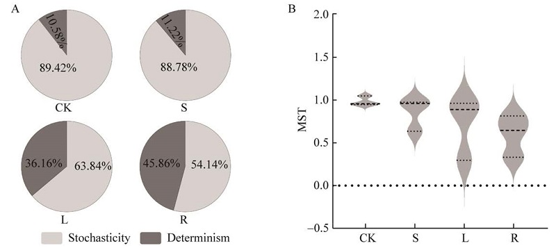
Shiro plays an essential role in the formation process of mycorrhizal edible fungi. Revealing the characteristics of microbial communities in the microhabitat of shiro is of great significance for understanding the formation mechanism of fruiting bodies of mycorrhizal edible fungi. In this study, four spatial gradient soil sample types were collected, including the soil at the base of fruiting bodies (R), surface soil around fruiting bodies (S), lower soil around fruiting bodies (L) and control soil (CK) in the non-fruiting area of Amanita sinensis. Using metagenomic sequencing technology, the fungal community diversity and assembly mechanism in the shiro soil of A. sinensis were analyzed. In total, 9 phyla, 51 classes, 137 orders, 372 families, 761 genera and 1 822 species of fungi were identified in the shiro soil. There were significant differences in species richness among fungal communities of different tested soil, showing R (1 653) > S (1 329) > L (1 307) > CK (968); there was no statistically significant difference in alpha diversity index among soil fungal communities in different spatial gradients of A. sinensis shiro. Ascomycota and Basidiomycota were predominant in the soil fungal communities, while Hypocreaceae was predominant family. NST and MST analysis showed that there were statistically significant discrepancies in the construction process of soil fungal community in different spatial gradients of A. sinensis shiro, and the proportion of deterministic process increased with the increase of soil depth and distance from the shiro. There were statistically significant discrepancies in soil fungal indicator species in different spatial gradients of A. sinensis, and the fungal indicator species in the soil at the base of fruiting bodies (R) were the most abundant. The results herein may serve as a theoretical reference for the artificial domestication and cultivation of A. sinensis.
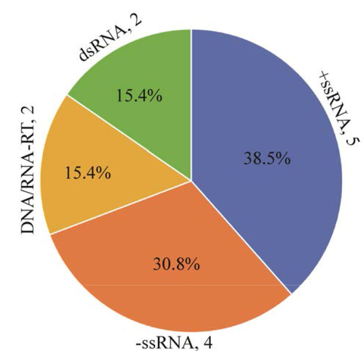
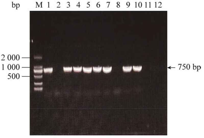
The diversity of mycoviruses in Fusarium oxysporum f. sp. niveum (FON) in Hebei Province is investigated. Screening of hypovirulent strains for providing biological control resources in the management of watermelon wilt was conducted on the basis of high-throughput sequencing and biological characterization of 130 FON strains in watermelon collected from Hebei Province. In total, 13 mycoviruses were identified, including five positive-sense single-stranded RNA viruses, four negative- sense single-stranded RNA viruses, two double-stranded RNA viruses, and two reverse-transcribing viruses. Among these, five viruses showed less than 70% similarity in their RdRp sequences to known viruses, suggesting they are potential new species. Strain Fo-66 harboring F. oxysporum f. sp. niveum Betachrysovirus 1 (FoNBV1) was focused. The genome of the virus consists of four dsRNA segments, with a total length of 12 574 bp. Compared with the virus-free strain Fo-66-VF, the mycovirus-infected strain Fo-66 exhibited 20.7% reduction in growth rate, 24.5% decrease in sporulation, and its pathogenicity was significantly reduced (disease index of watermelon decreased from 57.5 to 33.3). FoNBV1 was horizontally transmitted to F. graminearum and F. pseudograminearum via dual culture, and vertical transmission efficiency through conidia reached 100%. This study has revealed the diversity of mycoviruses in the pathogens causing Fusarium wilt of watermelon in Hebei Province. It is clarified that FoNBV1 can reduce the host's growth rate, spore production, and pathogenicity, and possesses hypovirulent properties, thereby providing a theoretical basis and candidate resources for the biological control of Fusarium wilt of watermelon.
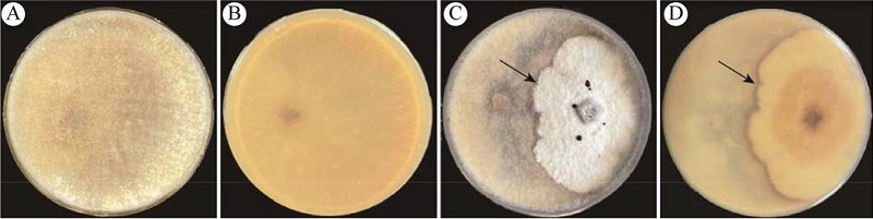
The pathogen of fungal diseases endangering Morchella sextelata in Beijing was isolated and purified by using tissue isolation method. Morphological and molecular biology methods were used for identification of the pathogen. Suitable growing temperatures and pH, and the effects of carbon and nitrogen sources on the growth rate, colony morphology, and sporulation of fungal hyphae were systematically analyzed. Through morphological observation and analysis of ITS, RPB2, and LSU gene sequences, the pathogen was identified as Pseudodiploospora longispora (similarity>99%). The Koch’s rule inoculation test verified the pathogenicity of the pathogen. It was confirmed that the optimal growth temperature of the fungus was 20 ℃, and the pH adaptation scope was wide, ranging from pH 5 to pH 9. The most suitable carbon source is sucrose, and the most suitable nitrogen source is malt extract powder. Under the conditions of 20 ℃ and pH 5, the highest sporulation was attained by using glucose as the carbon source and yeast extract as the nitrogen source. This test provides theoretical reference for preventing and controlling the white mold disease of cultivated Morchella sextelata.
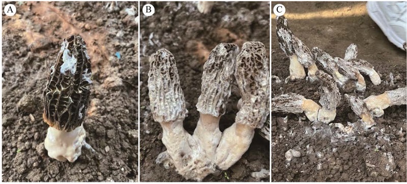
Morchella sextelata is an edible and medicinal fungus characterized by its preference for low temperature and high-humidity environments, and the species is rich in physiologically active substances such as amino acids, polysaccharides, and organic acids. With the expansion of artificial cultivation, disease problems in Morchella cultivation have been increasingly prominent, particularly white mold, rot, erythropathy, and cobweb disease. Among these, white mold disease, caused by Pseudodiploospora longispora, has emerged as the most serious and devastating fungal disease due to its wide occurrence and rapid spread. This study systematically investigated the infection process of P. longispora on Morchella mycelia and fruiting bodies using dual-culture assays, light microscopy, paraffin sectioning, and scanning electron microscopy (SEM). The results demonstrated that the pathogenic mycelia infected Morchella sextelata mycelia via adhesion, spiral coiling, and clip-like structures. Under conidial suspension concentration of ≥1×10³ spores/mL, mycelial growth of M. sextelata was significantly inhibited. The pathogen colonized the pileus surface, leading to tissue collapse and perforation as the infection progressed. This study revealed the pathological characteristics of P. longispora infecting mycelia and fruiting bodies of M. sextelata at different development stages, providing reference for the prevention and control of white mold disease in M. sextelata cultivation.
Cordyceps cateniannulata is an entomopathogenic fungus with significant biocontrol potential against agricultural and forestry pests. Although the transcriptome sequencing of this fungus has been completed, the lack of establishment of a genetic transformation system has severely hindered functional gene research. In this study, protoplasts of C. cateniannulata were successfully prepared and a PEG-mediated protoplast genetic transformation system was established, achieving stable expression of the exogenous GFP gene. Two novel hsp30 genes, Cchsp30a and Cchsp30b, were cloned from C. cateniannulata (GenBank accession numbers: PX352604 and PX352605, respectively). Using the established transformation system, overexpression mutant strains of Cchsp30a and Cchsp30b were obtained. Phenotypic analysis revealed that, compared to the wild-type strain, the mutant strains exhibited significant enhancement of spore germination rates under heat shock (48 °C), oxidative stress (menadione), salt stress (NaCl), and cell wall stress (Congo red), and enhanced germ tube elongation under UV radiation. These results confirm the positive regulatory role of Cchsp30 genes in broad-spectrum stress tolerance. Further transcriptome analysis under heat stress showed that a large number of differentially expressed genes (DEGs) in the mutants were significantly enriched in stress-related pathways, particularly the ABC transporter pathway. Notably, genes belonging to the ABCC transporter subfamily were most prominently differentially expressed in the mutants, suggesting that Hsp30 might enhance cellular stress adaptation by modulating the transcription of ABCC subfamily members, thereby influencing substance transport. In summary, this study not only established a genetic manipulation system for C. cateniannulata, but also but also preliminarily characterized the broad-spectrum stress resistance function of Cchsp30 and revealed its potential involvement in stress adaptation through regulating the transcription of ABCC subfamily transporters. These findings provide clues for further exploring the role of Cchsp30 in the stress resistance mechanisms of this entomopathogenic fungus.
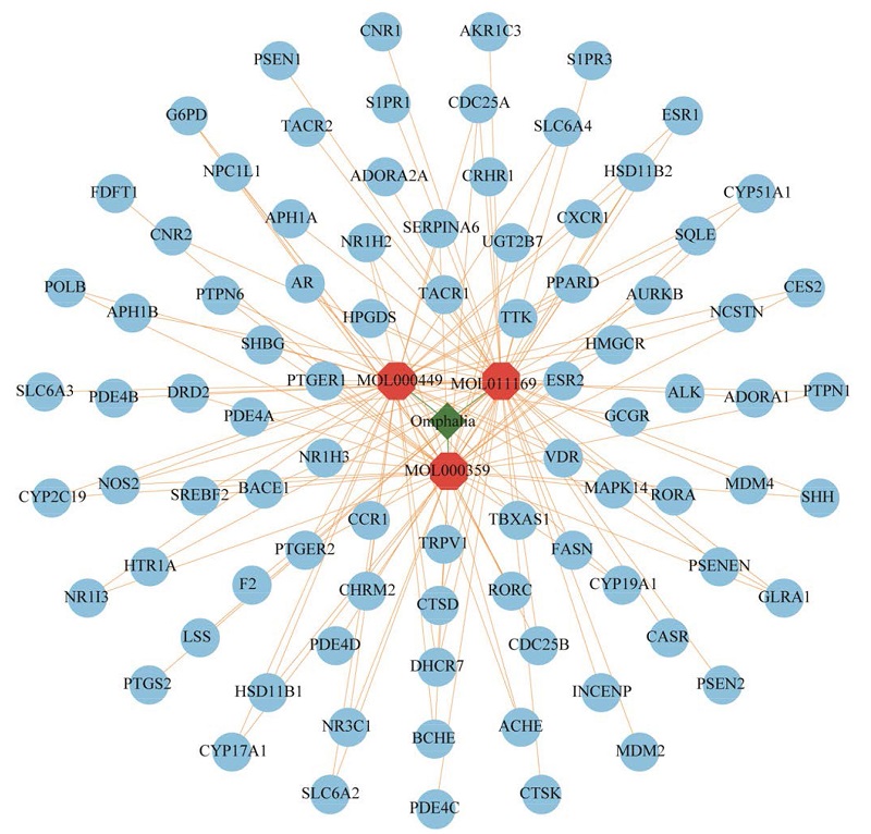
Epilepsy is a complex chronic neurological disorder, existing antiepileptic drug face limitations such as treatment resistance. Omphalia lapidescens sclerotium (lei wan, omphalia), a traditional fungal medicine, has demonstrated certain anticonvulsant effects, but its mechanisms remain unclear. This study aims at systematically elucidating the mechanism of omphalia in treating epilepsy through integrating network pharmacology, multi-omics Mendelian randomization (MR), and various bioinformatic approaches. Active ingredients of omphalia were screened using the TCMSP database, and their potential therapeutic targets were predicted. By combining MR analysis based on eQTL/pQTL data, differential gene expression analysis from microarray datasets and machine learning models, key targets for omphalia in epilepsy treatment were identified, and core targets were selected through topological analysis. Subsequently, GO/KEGG enrichment analysis, gene set enrichment analysis, immune infiltration assessment, molecular docking, and gut microbiota MR were applied to explore the functional mechanisms of omphalia. Three active ingredients, sitosterol, stigmasterol, and ergosterol peroxide, along with 87 potential targets were identified. Intersection with epilepsy-related targets obtained through various methods yielded seven key targets: RORC, S1PR3, ADORA1, PTGS2, RORA, CDC25B, and TBXAS1, among which PTGS2 and RORA were identified as core targets. Molecular docking showed binding energies below −11.0 kcal/mol for all active ingredients with core targets, indicating strong binding affinity. Enrichment analysis revealed that these targets are primarily involved in regulating lipid metabolism, circadian rhythm, inflammatory response, and sphingolipid signaling pathways. Immune infiltration analysis suggests that RORA activates CD8+ T cells and inhibits pro-inflammatory cells, while PTGS2 regulates dendritic cell activation. MR analysis of gut microbiota indicated causal associations between PTGS2/RORA and 17 microbial taxa including Bifidobacterium, suggesting omphalia might exert antiepileptic effects by modulating the gut-brain axis. This study reveals that omphalia exerts anticonvulsant effects through multi-target and multi-pathway synergistic regulation of immune inflammation, lipid metabolic homeostasis, circadian rhythm, and gut-brain interaction. The research overcomes the limitations of traditional network pharmacology and provides a reference for the clinical translation of omphalia and optimization of epilepsy treatment strategies.
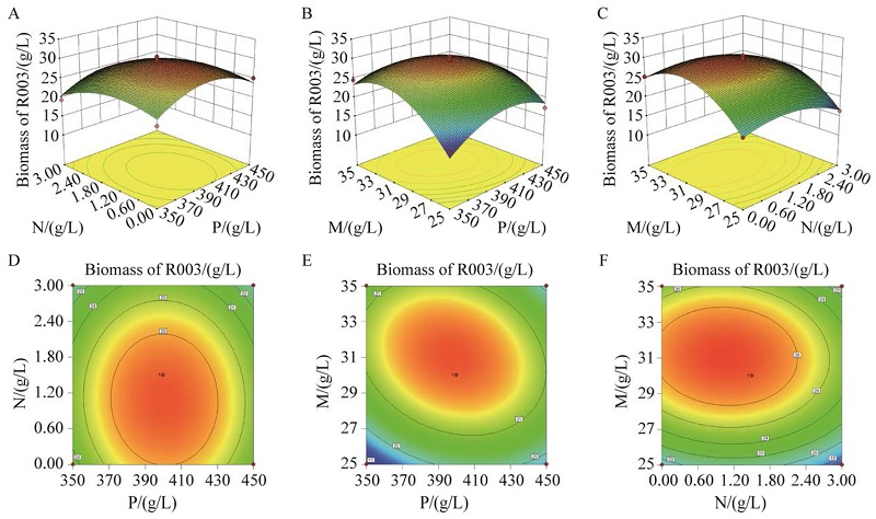
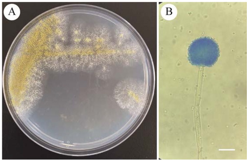
The wild strain 2019 MYKLR003 of Phlebopus roseus was used as the test material. Basic parameters for submerged culture were determined through single-factor experiments, and the medium formula was optimized using the response surface methodology. A high-activity induction formula was designed and verified, and the activity changes of peroxidase, laccase, and cellulase in mycelia under different fermentation conditions were determined. The results showed that this strain had a high oxygen demand for submerged culture and was highly sensitive to pH changes. The optimal pH value was 5 and liquid volume was 100 mL. The optimal medium formula (RA1) consisted of 432.35 g/L potato extract, 2.00 g/L tryptone, and 32.47 g/L mannitol. The actual measured mycelial biomass increased by 130.21% as compared with the predicted value. The addition of 100 μg/mL VB1, 50 μg/mL VB2, 50 μg/mL VB3, and 5 g/L carboxymethyl cellulose to RA1 significantly alleviated mycelial browning and led to regular and uniform mycelial morphology. Adjusting the fermentation endpoint to 18 days could achieve the maximum mycelial biomass synthesis while ensuring the simultaneous highest activities of the three extracellular enzymes. This study provides a research reference for the standardized production of high-activity submerged spawn of P. roseus.
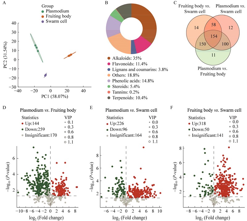
Physarum rigidum undergoes complex developmental changes during the plasmodial, fruiting body, and swarm cell stages. To elucidate the differences in metabolites and genes at different developmental stages, integrated analyses of metabolome and transcriptome were conducted. Based on metabolomics 499 stage-specific differential metabolites were identified, primarily comprising alkaloids, flavonoids, phenolics, steroids, and terpenoids. Pairwise comparisons revealed distinct metabolic profiles among the three stages: the plasmodial stage exhibited vigorous biosynthetic activity, with 226 metabolites upregulated as compared to the swarm cell stage; fruiting body stage exhibited upregulation of 259 and 318 metabolites as compared to the plasmodium and swarm cell stages, respectively. Transcriptomic analyses detected 35 844 differentially expressed genes. Functional enrichment analyses revealed highly active pathways of ribosome assembly and amino acid biosynthesis in the plasmodial stage, significant enrichment of upregulated genes in the MAPK signaling pathway and autophagy-related pathways in the fruiting body stage, and a characteristic maintenance of fundamental cellular activities such as endocytosis in the swarm cells. Integrated analyses revealed high consistency between metabolic and transcriptional changes in core metabolic pathways during plasmodium-involved transitions, including metabolic pathways, biosynthesis of secondary metabolites, carbon metabolism, and biosynthesis of amino acids; the fruiting body-to-swarm cell transition specifically involved cofactor biosynthesis and ABC transporters. This study provides the first multi-omics atlas of P. rigidum development, revealing stage-specific metabolic reprogramming predominantly driven by transcriptional regulation and identifying unique adaptive mechanisms at different developmental stages.
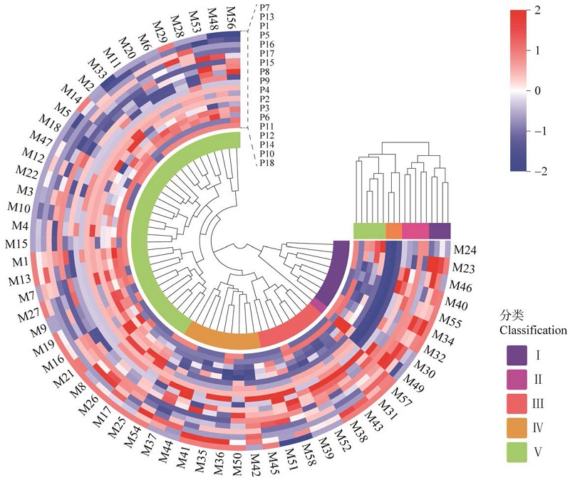
For the purposes of improving morel breeding efficiency, phenotypic diversity and variation level of morel germplasm resources of 58 strains preserved at the Sichuan Institute of Edible Fungi were analyzed. Eighteen phenotypic traits were tested according to DUS testing guidelines for Morchella, and comprehensive evaluations combined with genetic diversity, correlation analysis, cluster analysis, and principal component analysis were carried out. The results showed that among the 12 qualitative phenotypic traits, the genetic diversity index (Hʹ) of pileus ridge density was the highest (1.456), while that of the presence or absence of mycelial sclerotia was the lowest (0.432). For the 6 quantitative phenotypic traits, the Hʹ values ranged from 1.906 to 2.083, with an average of 1.974. Correlation analysis revealed that there were varied degrees of significant or extremely significant correlations among the 18 phenotypic traits. Cluster analysis divided the 58 germplasm resources into 5 groups, and each possessed typical phenotypic characteristics. Principal component analysis extracted 6 principal components with a cumulative contribution rate of 71.36%, separately corresponding to core dimensions such as pileus quality, mycelium evaluation, and ascocarp morphology. The comprehensive evaluation model constructed by principal components classified the germplasm into 6 groups, among which Morchella exuberans strain M49 had the highest comprehensive score (1.18). The prediction equation established by stepwise regression analysis (adjusted R2=0.931) indicated that 10 traits, including the distribution pattern of mycelial sclerotia and pigment intensity, were the key factors affecting the comprehensive performance of Morchella. This study confirmed that the phenotypic genetic diversity of morel germplasm was rich, and the constructed comprehensive evaluation system provided an efficient germplasm screening for genetic improvement and breeding of Morchella.
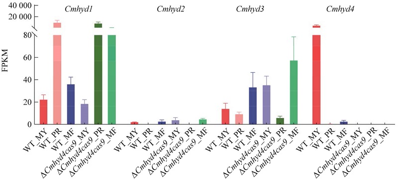
Hydrophobins, small secreted proteins unique to filamentous fungi, play critical roles in fungal growth and development yet their functional mechanisms are poorly understood. Previously, a hydrophobin gene Cmhyd4 knockout strain ∆Cmhyd4cas9 of Cordyceps militaris was generated via gene editing. A comparative analysis of mycelial growth and fruiting body development before and after Cmhyd4 deletion was conducted. It was revealed that the loss of Cmhyd4 promoted aerial hyphae growth, and increased the number of fruiting bodies and biological conversion efficiency per cultivation bottle. Using wild-type (WT) and ∆Cmhyd4cas9 strains as experiment materials, samples from the mycelia (MY), primordium (PR), and mature fruiting body (MF) were subjected to comparative transcriptomic analysis. The results showed that Cmhyd4 deletion altered gene transcript levels across all stages as compared with WT strain. The highest differentially expressed genes (DEGs) occurred at PR stage (349), and MY stage (179) was the next, while the fewest at MF stage (93). The transcriptional patterns of hydrophobin family genes at different developmental stages provided critical insights into their functions. The deletion of Cmhyd4 altered the transcription levels of the other family members, demonstrating significant gene compensation effects. GO enrichment analysis revealed that after Cmhyd4 gene deletion, DEGs at the MY stage were primarily associated with cell cycle and mitosis, whereas DEGs at PR and MF stages were linked to abiotic stress response. Consisted with GO analysis, the ∆Cmhyd4cas9 strain exhibited significant enhancement of NaCl stress resistance in fruiting bodies, while no notable changes were observed in the mycelial stage. RT-qPCR validation confirmed that four genes in the osmotic stress response pathway showed no differential expression at MY stage after Cmhyd4 gene deletion, but significant upregulation occurred in PR and MF stages. This study comprehensively elucidates the impacts of Cmhyd4 on the growth and development of C. militaris, along with its regulatory role in gene expression, providing genetic resources for future molecular breeding of C. militaris.
Aspergillus flavus widely presents in crop seeds and its metabolite aflatoxin B1 (AFB1) poses threats to food security and human health. As a broad-spectrum antibacterial substance, bile acids can exert an inhibitory effect on A. flavus HL2, a high AFB1-producing strain isolated from corn. Among them, chenodeoxycholic acid (CDCA), which is more hydrophobic, inhibits the growth of A. flavus HL2 by destroying cell membrane homeostasis and releasing intracellular K+, with a MIC of 0.2 mg/mL. Hyodeoxycholic acid (HDCA), on the other hand, has a stronger inhibitory effect on AFB1 synthesis. By inhibiting the expression of AFB1 synthesis-related genes, 29.85% AFB1 synthesis can be reduced at a concentration of 0.5×MIC. Therefore, the combination of different bile acids provides a theoretical basis for the development of biological antifungal agents. At the same time, bile acids can combine with AFB1 to form a co-precipitation, which has the effect of removing AFB1. Among them, chenodeoxycholic acid can reduce AFB1 content by 55.55%. Bile acids possess both the functions of antifungal agents and mycotoxin binders, laying a foundation for their application in mold prevention and control in grain and animal feed products.
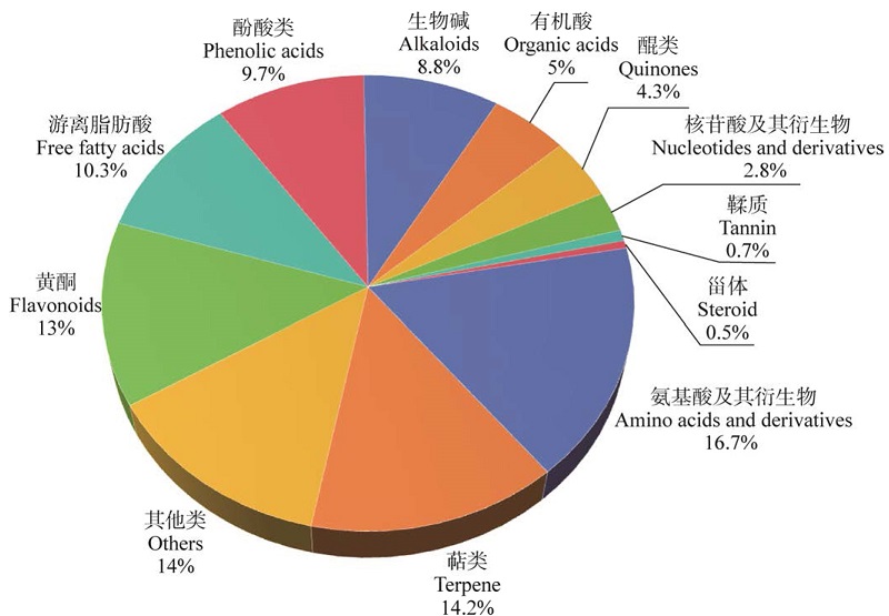
Antioxidant and antibacterial properties of the acetone-extracted preparation of Sticta nylanderiana (SE) were evaluated. Antioxidant activity assessed via DPPH, ABTS, and hydroxyl radical scavenging assays revealed that SE could significantly scavenged DPPH and ABTS+ free radical, with IC₅₀ values of (71.69±0.43) μg/mL and (172.7±3.01) μg/mL, respectively. Antibacterial activity determined through filter paper disc diffusion and broth dilution methods demonstrated the inhibitory effects of SE against three common bacterial strains. The minimum inhibitory concentration (MIC) values were 0.625 mg/mL for Bacillus subtilis, 0.625 mg/mL for Escherichia coli, and 1.25 mg/mL for Staphylococcus aureus. Chemical profiling using UPLC-MS/MS identified a total of 2 171 metabolites, including amino acids and their derivatives, terpenoids, flavonoids, and phenolic acids. These findings indicate that SE possesses notable antioxidant and antibacterial activities, underscoring its potential for pharmaceutical and health-related applications. This study provides a scientific foundation for further exploration and utilization of bioactive compounds in lichens.
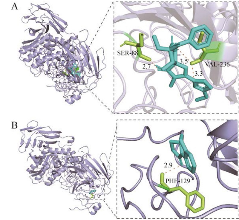
The secondary metabolites of Aspergillus fumigatus ZNM2-3 isolated from the sediment of Nali Karst Cave in Shangsi County, Fangchenggang, Guangxi were studied. The secondary metabolites were isolated and purified by normal phase silica gel column chromatography and high-performance liquid chromatography. The structures were identified by electrospray ionization mass spectrometry (ESI-MS), NMR and other methods. The cytotoxic activity and α-glucosidase inhibitory activity of the compounds were determined, and the active compounds were verified by molecular docking. In total, 13 compounds were isolated from Aspergillus fumigatus ZNM2-3. Of which compound 6 had cytotoxic activity; compounds 8 and 12 had certain inhibitory effects on α-glucosidase, with IC50 values of 9.46 μmol/L and 10.17 μmol/L, respectively. Molecular docking results showed that both of these two compounds could bind α-glucosidase in the form of hydrogen bonds, thereby inhibiting the degradation of the substrate by the enzyme.