


灵芝Ganoderma lingzhi Sheng H. Wu et al.是一种著名的药用真菌(戴玉成等 2021),具有较高的药用价值,是我国传统的名贵中药材。灵芝中富含多种功能成分,其中包括灵芝多糖(Seweryn et al. 2021)、灵芝三萜(罗云等 2021)、氨基酸(Sliva 2004)、核苷(刘艳芳等 2021)、蛋白质(周选围等 2007)和微量元素(Singh et al. 2020)等。β-葡聚糖作为灵芝中主要的活性成分,在免疫调节(Koliakos et al. 2021;刘静等 2021)、抗肿瘤(Mo et al. 2017)、抗炎(Abreu et al. 2019)及抗氧化(徐勇亮和徐军伟 2022)等方面有较好的活性。前期实验室从灵芝子实体中纯化出一种β-葡聚糖组分GLP20 (Liu et al. 2014),其分子量为2.42×10³ kDa,是以β-(1→3)-葡萄糖连接为主链、β-(1→6)-葡萄糖连接为支链的葡聚糖,且主链与支链的比例为3:1。研究发现该葡聚糖具有较好的生物活性,然而由于其分子量大、黏度大、溶解性相对较差等特点影响了其进一步的开发利用。相关研究表明多糖降解为低聚糖不仅可以降低分子量,提高其水溶性,还可以保留甚至提高多糖原有的生物活性(Kim et al. 2005;Yan et al. 2016)。还有研究表明,寡糖的活性取决于它们的结构,包括分子量、聚合度(DPs)和修饰等(Iwamoto et al. 2005;Chen et al. 2017;Kaczmarek et al. 2019)。在Shi et al. (2018)的研究中,使用葡聚糖酶将线性β-(1→3)-葡萄糖连接的可德兰多糖(curdlan)降解,得到了DP2-5的β-(1→3)-葡寡糖,并检测其益生元效应,结果表明,聚合度为2和3的β-(1→3)-葡寡糖可以发挥更好的益生元作用。由于灵芝β-葡聚糖GLP20组分结构中连有β-(1→6)-葡萄糖支链,因此降解得到的β-葡寡糖结构会更丰富。本实验室前期通过微波法将灵芝β-葡聚糖GLP20降解为低聚糖片段,初步分离后得到具有抗炎活性的葡寡糖组分GLPW-A (DP2-14) (秦秀 2021)。为了进一步明确其中发挥主要抗炎活性的寡糖片段,本研究采用Bio-Gel P-2凝胶柱对灵芝β-葡寡糖GLPW-A组分进行分离纯化,分别从洗脱液的选择、流速以及每管接样量3个方面对分离效果进行优化,得到最优的分离条件,然后通过MALDI-TOF-MS系统对分离得到的组分进行结构解析,并在THP-1型巨噬细胞炎症模型中评价各组分的抗炎活性,比较在一定浓度下不同聚合度葡寡糖的抗炎效果,为葡寡糖的构效关系研究提供基础。
1 材料与方法
1.1 供试材料
1.1.1 主要试剂
β-葡寡糖GLPW-A制备:参考秦秀(2021)的方法,将10 mg/mL的灵芝β-葡聚糖GLP20组分置于微波消解罐中,设定微波功率为1 000 W,温度为140 ℃,处理60 min得到降解产物GLPW,然后将30 mg/mL的GLPW使用90%乙醇溶液沉淀4 h,取醇沉后上清冻干得到β-葡寡糖GLPW-A组分(DP2-DP14);人单核细胞系(THP-1) (中国科学院上海细胞库);RPMI-1640培养基和胎牛血清(Gibco公司);Human TNF-α ELISA试剂盒(北京四正柏生物公司);脂多糖(LPS)、Alamar Blue试剂、佛波酯(PMA)和β-巯基乙醇(Sigma公司);青霉素和链霉素(Amresco公司);葡萄糖(DP1,国药集团化学试剂有限公司);昆布二糖(DP2)、昆布三糖(DP3)、昆布四糖(DP4)、昆布五糖(DP5)和昆布六糖(DP6) (Megazyme公司);其他试剂均为国产分析纯。
1.1.2 仪器和设备
高通量密闭高压微波消解仪(CEM公司);Dionex ICS-5000+离子色谱系统(Dionex公司);MALDI-TOF-MS (AB SCIEX公司);AKTA中压层析系统(GE公司);分析天平(Mettler Toledo公司);CO2培养箱(Thermo Fisher公司)。
1.2 方法
1.2.1 凝胶色谱分离条件优化
准确称取25 mg GLPW-A组分溶于0.5 mL超纯水中,经0.45 μm滤膜过滤后,采用Bio-Gel P-2凝胶柱分离。分别从洗脱液种类、流速及每管接样量3个方面进行优化,每管多糖吸光值采用苯酚-硫酸法(张剑等 2017)测定,并以洗脱体积或管号为横坐标、吸光值为纵坐标制作洗脱曲线图,根据洗脱曲线收集各洗脱组分。
1.2.2 HPAEC-PAD系统分析
准确称取2 mg待测样品溶于1.0 mL超纯水中,12 000 r/min离心10 min后过0.45 μm滤膜,参考Catenza & Donkor (2021)的报道,采用高效阴离子交换色谱联用脉冲安培检测器(HPAEC- PAD)分析寡糖的聚合度。色谱柱:CarbopacTM PA-100柱(4 mm×250 mm,Dionex公司);梯度洗脱方式:流动相A:150 mmol/L NaOH,流动相B:150 mmol/L NaOH和500 mmol/L CH3COONa;流速:1 mL/min;柱温和脉冲安培检测器的温度维持在30 ℃;进样量:25 μL。梯度洗脱程序如下:0-5 min,A/B为90/10 (体积比);5-30 min,A/B为80/20 (体积比);30-30.1 min,A/B为30/70 (体积比);30.1-40 min,A/B为0/100 (体积比);40-45 min,A/B为90/10 (体积比)。
1.2.3 MALDI-TOF-MS系统分析
以基质辅助激光解析电离飞行时间质谱法(MALDI-TOF-MS)对分离得到的组分的聚合度进行分析。精确称取5 mg待测样品溶于1 mL超纯水中,取1 μL待测液与1 μL DHB基质充分混合后,吸取1 μL点样于样品板指定位置,室温下待溶剂挥发后备用。质谱条件:正离子模式;DHB基质:DHB (2,5-二羟基苯甲酸)溶于甲醇/水溶液中(50/50,体积比,含0.1%的三氟乙酸)配制成10 mg/mL的DHB基质溶液;扫描分子量范围:200-4 000 g/mol。
1.2.4 产物的抗炎活性评价
样品的配制:精确称取5 mg待测样品置于已灭菌的离心管中,加入1 mL的无菌水,充分溶解后经0.22 μm的无菌水相滤膜过滤除菌备用。
细胞培养:THP-1细胞用含10%胎牛血清、1%青霉素、1%链霉素和0.5% β-巯基乙醇的RPMI-1640培养基于37 ℃、5% CO2培养箱中孵育,一周换液2次。
抗炎活性测定:取对数生长期的THP-1细胞,将细胞以5×10⁵ 个/mL的密度接种到96孔板中,加入PMA (终浓度为30 ng/mL)培养40 h,待THP-1被诱导为巨噬细胞后弃去上清,每孔加入160 μL的RPMI-1640培养基,空白对照组加入40 μL的磷酸盐缓冲溶液(PBS,含0.14 mol/L NaCl、0.002 7 mol/L KCl、0.004 mol/L Na2HPO4、0.002 mol/L KH2PO4,pH 7.4),阳性对照组加入20 μL的LPS (作用浓度为50 ng/mL)和20 μL的PBS,实验组加入20 μL的LPS (作用浓度为50 ng/mL)和20 μL不同浓度的样品液。于5% CO2培养箱中37 ℃培养24 h后取上清,按照ELISA细胞因子试剂盒说明书测定TNF-α释放量。
2 结果与分析
2.1 凝胶柱分离寡糖组分的条件优化
为了优化凝胶柱层析分离葡寡糖的条件,分别考察了洗脱液种类、流速以及每管接样量3个方面对分离效果的影响。
2.1.1 洗脱液对分离效果的影响
分别选用超纯水和NH4HCO3为洗脱液,其分离效果见图1,结果发现以超纯水为洗脱液时,各组分不能达到有效分离。以0.1 mol/L的NH4HCO3为洗脱液时,样品可被分离为4个峰,分离效果优于超纯水,这可能是由于以NH4HCO3为洗脱液时可减小寡糖与色谱柱之间的吸附力,使样品可根据分子量的不同大小进行分离,因此选用以0.1 mol/L的NH4HCO3为洗脱液。
图1
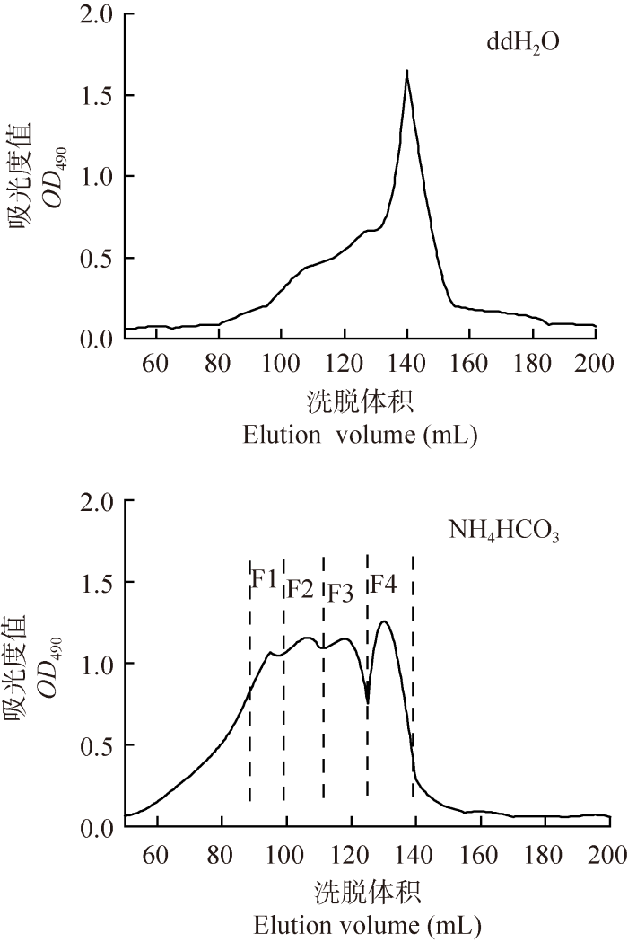
图1
不同洗脱液对寡糖分离效果的影响
Fig. 1
Effects of different eluents on the separation of oligosaccharides from Ganoderma lingzhi.
2.1.2 洗脱流速对分离效果的影响
考察了洗脱流速为0.05、0.1和0.3 mL/min时凝胶色谱柱对组分GLPW-A的分离效果(图2)。在洗脱流速为0.05-0.3 mL/min下,随着洗脱流速的降低,各洗脱峰之间的分离度逐渐提高。考虑到流速为0.05 mL/min与0.1 mL/min时对寡糖分离效果的影响差别较小,仅在洗脱体积为55-80 mL处表现出差异,综合考虑,确定以0.1 mL/min的流速进行分离。
图2
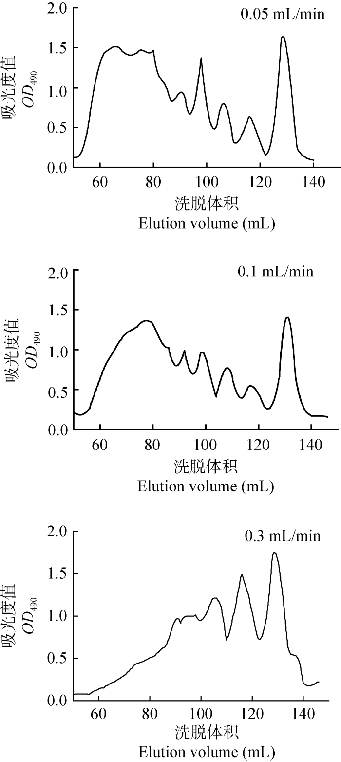
图2
不同洗脱流速对寡糖分离效果的影响
Fig. 2
Effects of elution flow rates on the separation of oligosaccharides from Ganoderma lingzhi.
2.1.3 每管接样量对分离效果的影响
在选择了合适的洗脱液和流速后,进一步考察了接样体积对分离效果的影响(图3)。2 mL/管的接样量避免了各组分之间的交叉,接样体积为2 mL/管时比接样体积为5 mL/管时分离效果更好。
图3
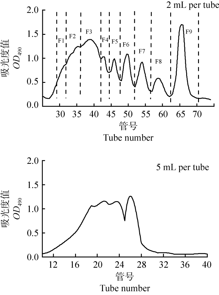
图3
每管接样量对寡糖分离效果的影响
Fig. 3
Effects of collection volume per tube on the separation of oligosaccharides from Ganoderma lingzhi.
图4
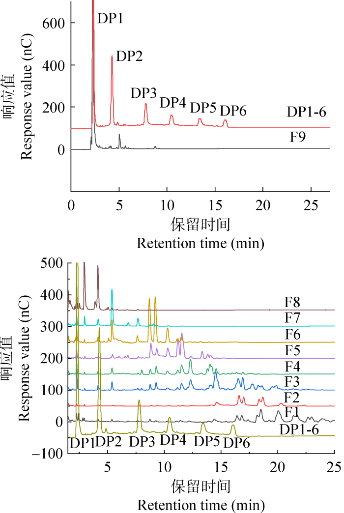
图4
凝胶分离组分的高效阴离子色谱图
Fig. 4
HPAEC analysis of oligosaccharide fractions separated by gel chromatography.
2.2 HPAEC-PAD系统分析组分的组成
为明确各峰之间的分离效果,采用HPAEC对收集到的8个组分和标准品(DP1-DP6)进行分析(图4)。葡萄糖标准品和DP2-DP6的β-(1→3)-葡寡糖标准品出峰时间分别为:2.3 min (葡萄糖)、4.3 min (DP2)、7.8 min (DP3)、10.5 min (DP4)、13.4 min (DP5)和16.0 min (DP6)。将分离得到的8个组分和寡糖标准品的出峰时间对比,发现除上述出峰时间外还有其他峰出现。在Xie et al. (2021)的报道中,使用HPAEC-PAD-MS平台分析葡聚糖酶解产物的聚合度变化,并用MS/MS法确定了寡糖中各糖苷键的连接方式,发现同一聚合度葡寡糖出峰时间不同,且MS/MS分析结果证明其连接方式不同。F8组分除了在4.3 min处有一个峰(图4)外,3 min处还有一个峰出现,表明该组分主要含有连接方式不同的二糖;F7组分的色谱图中主要为5.3 min的峰,表明产物中主要含有三糖;而F6组分的HPAEC图谱中存在2个相近的峰,保留时间分别为8.7 min和9.2 min,此外在10.5 min处还存在一个较小的峰,推测F6组分同时存在多个聚合度的寡糖;F4与F5组分的出峰时间相似,但F4在14.0 min处的色谱峰响应值明显高于F5,表明F4与F5组分并未达到较好的分离效果;而F1组分主要为保留时间为16.3-25.2 min的聚合度较大的寡糖片段。结合文献推测灵芝β-葡聚糖降解得到的产物中葡寡糖种类更丰富。
2.3 MALDI-TOF-MS系统测定组分的聚合度
图5
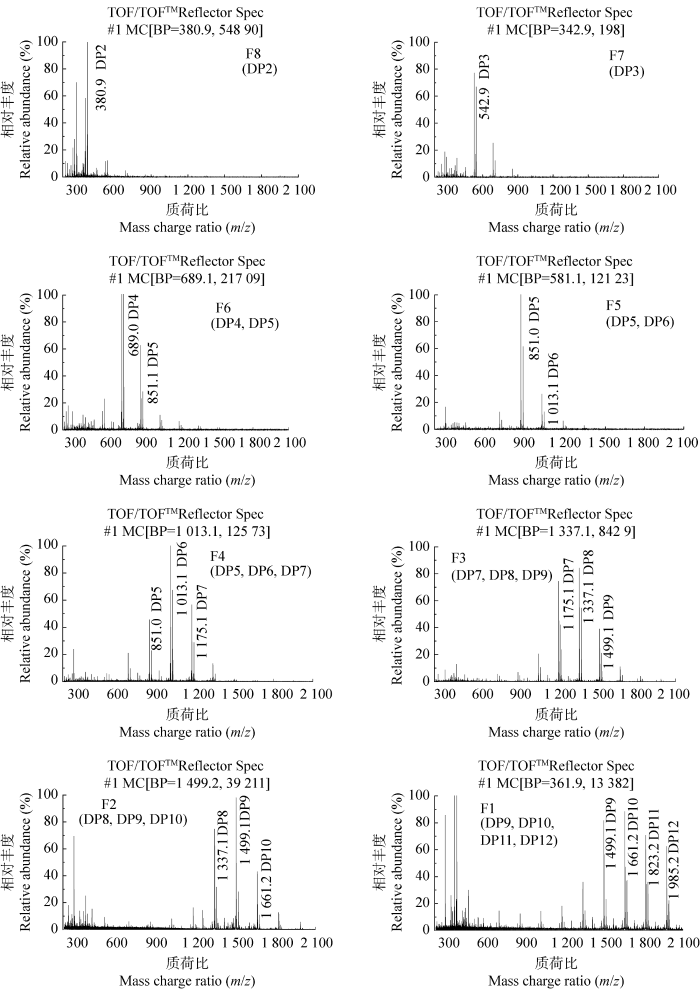
图5
凝胶分离各组分的MALDI-TOF-MS质谱图
Fig. 5
MALDI-TOF-MS mass spectrum of oligosaccharide fractions separated by gel chromatography.
表1 凝胶分离各组分的质谱结果
Table 1
| 样品 Sample | 质荷比 m/z | 聚合度 Polymerization degree |
|---|---|---|
| F1 | 1 499.1, 1 661.2, 1 823.2, 1 985.2 | DP9, DP10, DP11, DP12 |
| F2 | 1 337.1, 1 499.1, 1 661.2 | DP8, DP9, DP10 |
| F3 | 1 175.1, 1 337.1, 1 499.1 | DP7, DP8, DP9 |
| F4 | 851.0, 1 013.1, 1 175.1 | DP5, DP6, DP7 |
| F5 | 851.0, 1 013.1 | DP5, DP6 |
| F6 | 689.0, 851.1 | DP4, DP5 |
| F7 | 542.9 | DP3 |
| F8 | 380.9 | DP2 |
DP:寡糖聚合度
DP: Degree of polymerization of oligosaccharides.
2.4 凝胶分离产物的抗炎活性评价
肿瘤坏死因子-α (tumor necrosis factor, TNF-α)是一种促炎细胞因子,在炎症条件下TNF-α水平显著升高(黄嘉欢等 2022)。为探讨各聚合度寡糖组分的抗炎活性,分别测定各组分在LPS刺激下THP-1型巨噬细胞培养液中的TNF-α水平。其中,由于F2组分的糖含量较低,F4与F5组分所含的寡糖聚合度基本相同,所以选择F1、F3、F5、F6、F7和F8共6个组分进行抗炎活性研究。与LPS处理的细胞相比,分离得到的葡寡糖组分能显著降低TNF-α的释放水平(图6),说明对TNF-α的释放具有抑制作用,具有抗炎活性。且分离后的组分较原样GLPW-A的抗炎效果总体提高,尤其是在作用浓度为10 μg/mL时,与阳性对照组相比,F7 (DP3)组分表现出极显著的抗炎活性(P<0.01);在0.5 μg/mL作用浓度下,高聚合度葡寡糖组分F1、F3以及F8 (DP2)组分均与阳性对照LPS组表现出显著性差异(P<0.05);在作用浓度为0.5-10 μg/mL范围内,F5、F6组分未表现出抗炎活性,这2个组分分别主要含有四糖、五糖和五糖、六糖。综上推测,灵芝β-葡聚糖微波降解后产物的抗炎活性可能与寡糖聚合度有关。
图6
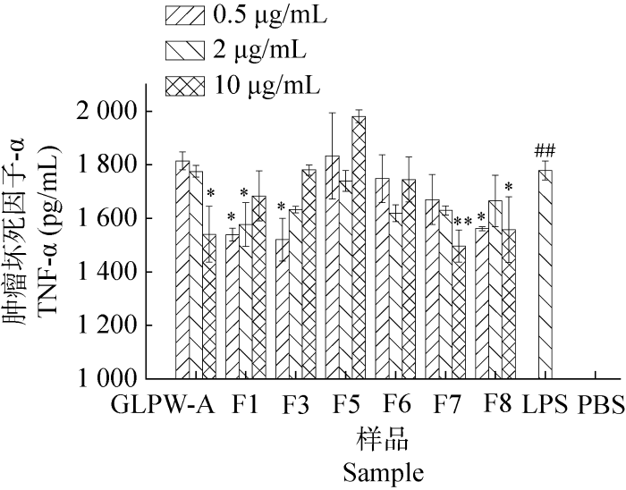
图6
不同样品对THP-1型巨噬细胞分泌TNF-α细胞因子的影响
##代表LPS组数据与阴性对照PBS组在P<0.01水平下具有显著性差异,*代表数据与阴性对照无菌水组在P<0.05水平下具有显著性差异,**代表数据与阳性对照LPS组在P<0.01水平下具有显著性差异
Fig. 6
Effects of different samples on the production of TNF-α cytokine released by THP-1 macrophages.
##P<0.01 compared to the negative control, *P<0.05, **P<0.01 compared to the positive control.
3 讨论
对灵芝葡聚糖降解后得到的一系列葡寡糖产物进行分离纯化,可为后续寡糖片段的结构鉴定和活性研究奠定基础。由于柱层析法具有选择性好、分离效率高、操作简便等优点,被广泛应用于糖类物质的分离纯化过程。姜瑞芝等(2008)使用Sephadex G-25分离猴头菌寡糖得到HEP-1-1、HEP-1-2和HEP-1-3,其中HEP-1-3是由二糖-七糖组成的混合葡寡糖。柱层析法通常通过调整流速、流动相、洗脱速度或优化柱长、柱温等影响分离效果的因素,来提高寡糖分离纯化的效果。琚洋洋(2016)采用凝胶排阻色谱Bio-Gel P2对壳寡糖进行分离纯化,以纯化出更多的壳寡糖单体为目标,对上样量和洗脱流速进行考察,分离纯化得到4个壳寡糖单体:壳二糖、壳三糖、壳四糖和壳五糖。高敏(2021)在对籽瓜果胶低聚糖采用Sephadex G-25分离纯化时,分别对洗脱液、流速及柱长进行优化,分离得到3个低聚糖组分。本研究采用Bio-Gel P-2凝胶柱对GLPW-A组分分离,分别从洗脱液、流速和每管接样量这3个洗脱条件进行优化,当洗脱条件为洗脱液为0.1 mol/L的NH4HCO3溶液,流速为0.1 mL/min,洗脱过程中每2 mL为一管进行收集时,分离效果最好,且分离得到8个组分。并通过阴离子交换色谱法和质谱法对富集得到的8个组分的聚合度分布进行了表征,各组分的聚合度分别为:F1:DP9-DP12,F2:DP8-DP10,F3:DP7-DP9,F4:DP5-DP7,F5:DP5-DP6,F6:DP4-DP5,F7:DP3,F8:DP2,表明通过该分离方法实现了对灵芝β-葡寡糖的初步分离。
已有研究表明,葡寡糖的聚合度与其生物活性密不可分。香菇β-葡聚糖作为我国抗肿瘤和抗肝炎病毒的临床一线用药,宁君和孔繁祚(2001)成功合成以β-(1→6)、β-(1→3)糖苷键连接的香菇葡寡糖片段,发现其可以激活人体的免疫系统产生免疫应答进而杀死肿瘤细胞,具有很好的抗肿瘤作用。Gissibl et al. (2018)使用葡聚糖酶处理微波预降解的裸藻β-(1→3)-葡聚糖得到DP2-DP7和DP2-DP59的葡寡糖,并证明低聚葡寡糖(DP2-DP7)相比于高聚葡寡糖(DP2-DP59)促进巨噬细胞释放TNF-α的水平更高,免疫活性显著增强。本研究的抗炎活性实验表明,与LPS处理的细胞相比,分离得到的葡寡糖组分能显著降低TNF-α的水平,且分离后的组分较原样GLPW-A的抗炎效果总体提高,其中,在作用浓度0.5-10 μg/mL范围内,部分组分与阳性对照LPS组相比表现出显著性差异,但含五糖、六糖的F5组分以及含有四糖、五糖的F6组分在该浓度范围内未表现出抗炎活性,因此推测样品的抗炎活性与寡糖片段的聚合度有关。为进一步明确寡糖的构效关系,将在后期对降解产物进行进一步的分离纯化,以期得到单一聚合度的寡糖并对其进行结构鉴定,以便为不同聚合度以及连接方式的寡糖的定量、定性分析及活性评价提供研究基础。
参考文献
Gelling functional property, anti-inflammatory and antinociceptive bioactivities of β-D-glucan from the edible mushroom Pholiota nameko
DOI:10.1016/j.ijbiomac.2018.09.062 URL [本文引用: 1]
Recent approaches for the quantitative analysis of functional oligosaccharides used in the food industry: a review
DOI:10.1016/j.foodchem.2021.129416 URL [本文引用: 1]
Alginate oligosaccharide DP 5 exhibits antitumor effects in osteosarcoma patients following surgery
DOI:10.3389/fphar.2017.00623 URL [本文引用: 1]
Diversity and systematics of the important macrofungi in Chinese forests
The separation, analysis and antibacterial mechanism of seed melon pectin oligosaccharide
Microwave pretreatment of paramylon enhances the enzymatic production of soluble β-1,3-glucans with immunostimulatory activity
DOI:S0144-8617(18)30570-8
PMID:29891305
[本文引用: 1]

A hydrothermal microwave pretreatment was established to facilitate the enzymatic production of soluble bioactive β-1,3-glucans from the recalcitrant substrate paramylon. The efficacy of this pretreatment was monitored with a newly developed direct Congo Red dye-based assay over a range of temperatures. Microwave pretreatment at 170 °C for 2 min resulted in a significantly enhanced enzymatic hydrolysis of paramylon. The action of endo-β-1,3- and exo- β-1,3-glucanases on the microwave-pretreated paramylon produced soluble β-1,3-glucans with degrees of polymerisation (DP) ranging from 2-59 and 2-7, respectively. In comparison, acid-mediated hydrolysis of untreated paramylon resulted in β-1,3-glucans with a DP range of 2-38. The hydrolysates were assayed on their immunostimulatory effect on murine macrophages by measuring the production of the inflammation-linked marker tumour necrosis factor alpha (TNFα) using immunofluorescence. All of the tested hydrolysis products were shown to induce TNFα production, with the most significant immunostimulatory effect observed with the hydrolysate from the exo-β-1,3-glucanase treatment.Copyright © 2018 Elsevier Ltd. All rights reserved.
Research progress on the preparation and anti-inflammatory mechanism of oligosaccharides
Structure-activity relationship of alginate oligosaccharides in the induction of cytokine production from RAW264. 7 cells
DOI:10.1016/j.febslet.2005.07.007
PMID:16055120
[本文引用: 1]
Guluronate and mannuronate oligomers with various degree of polymerization were prepared from polyguluronate (PG) and polymannuronate (PM) with an alginate lyase from a Pseudoalteromonas sp., and their activities to induce cytokine secretion from mouse macrophage cell line RAW264.7 cells were examined. Enzymatically depolymerized unsaturated alginate oligomers induced tumor necrosis factor (TNF)-alpha secretion from RAW264.7 cells in a structure-depending manner, while the activities of saturated alginate oligomers prepared by acid hydrolysis were fairly low or only trace levels. These results suggest that unsaturated end-structure of alginate oligomers was important for the TNF-alpha-inducing activity. Among the unsaturated guluronate (G3-G9) and mannuronate (M3-M9) oligomers, G8 and M7 showed the most potent activity, respectively. Bio-Plex assay revealed that interleukin (IL)-1alpha, IL-1beta, and IL-6 secretion from RAW264.7 cells were also induced by unsaturated alginate oligomers with similar structure-activity relationship profiles as seen in TNF-alpha, and the most potent activities were observed with G8 and M7. These results suggest that G8 and M7 may have the most suitable molecular size or entire structural conformation as stimulant for cytokine secretion. Since antibodies to Toll-like receptor (TLR)2 and TLR4 effectively inhibited the G8- and M7-induced production of TNF-alpha, these alginate oligomers may stimulate innate immunity through the pattern recognition receptors on macrophages similar to microbial products.
Chemical study on oligosaccharides of Hericium erinaceus
Preparation, purification and bioactivities of chitooligosaccharides
Enzymatic modifications of chitin, chitosan, and chitooligosaccharides
DOI:10.3389/fbioe.2019.00243
PMID:31612131
[本文引用: 1]
Chitin and its N-deacetylated derivative chitosan are two biological polymers that have found numerous applications in recent years, but their further deployment suffers from limitations in obtaining a defined structure of the polymers using traditional conversion methods. The disadvantages of the currently used industrial methods of chitosan manufacturing and the increasing demand for a broad range of novel chitosan oligosaccharides (COS) with a fully defined architecture increase interest in chitin and chitosan-modifying enzymes. Enzymes such as chitinases, chitosanases, chitin deacetylases, and recently discovered lytic polysaccharide monooxygenases had attracted considerable interest in recent years. These proteins are already useful tools toward the biotechnological transformation of chitin into chitosan and chitooligosaccharides, especially when a controlled non-degradative and well-defined process is required. This review describes traditional and novel enzymatic methods of modification of chitin and its derivatives. Recent advances in chitin processing, discovery of increasing number of new, well-characterized enzymes and development of genetic engineering methods result in rapid expansion of the field. Enzymatic modification of chitin and chitosan may soon become competitive to conventional conversion methods.Copyright © 2019 Kaczmarek, Struszczyk-Swita, Li, Szczęsna-Antczak and Daroch.
Anti-diabetic activity of beta-glucans and their enzymatically hydrolyzed oligosaccharides from Agaricus blazei
DOI:10.1007/s10529-005-2225-8 URL [本文引用: 1]
Immunomodulation through beta-D-glucan in chemically-induced necrotizing pancreatitis
DOI:10.1016/j.jss.2020.12.020
PMID:33421796
[本文引用: 1]
Although the ability of β-D-glucan and monophosphoryl lipid A (MPLA) to modulate immune responses has been studied in human primary cells, their effect on sterile inflammation models such as necrotizing pancreatitis has never been investigated.85 male New Zealand rabbits were assigned into following groups: A: control, B: pretreatment with β-D-glucan 3 d before pancreatitis, C: pretreatment with MPLA 3 d before pancreatitis, D: pretreatment with β-D-glucan and laminarin 3 d before pancreatitis, E: treatment with β-D-glucan 1 d after pancreatitis, and F: MPLA 1 d after pancreatitis. Pancreatitis was induced by sodium taurocholate injection into the pancreatic duct and parenchyma. Survival was recorded for 21 d. On days 1, 3, and 7, blood was collected for amylase measurement. Peripheral blood mononuclear cells were isolated and stimulated for tumor necrosis factor alpha and interleukin 10 production. Pancreatic necrosis and tissue bacterial load were assessed.21-d survival was prolonged after pretreatment or treatment with β-D-glucan; this benefit was lost with laminarin administration. At sacrifice, pancreatic inflammatory alterations were more prominent in the control group. Bacterial load was lower after pretreatment or treatment with β-D-glucan and MPLA. Tumor necrosis factor alpha production from stimulated peripheral blood mononuclear cells was significantly decreased, whereas interleukin 10 production remained unaltered after pretreatment or treatment with β-D- glucan.β-D-glucan reduces mortality of experimental pancreatitis in vivo. This is mediated through attenuation of cytokine production and prevention of bacterial translocation.Copyright © 2020 Elsevier Inc. All rights reserved.
Characterization and dectin-1 activation activity of polysaccharides from fruiting bodies of five edible fungi
Comparison of bioactive components in fruiting body and fermented mycelium of Ganoderma leucocontextum
Physicochemical characterization of a high molecular weight bioactive β-D-glucan from the fruiting bodies of Ganoderma lucidum
DOI:10.1016/j.carbpol.2013.10.024 URL [本文引用: 1]
Research progress on pharmacological activities of triterpenes from Ganoderma lucidum
Anti-tumor effects of (1→3)-β-D-glucan from Saccharomyces cerevisiae in S180 tumor-bearing mice
DOI:S0141-8130(16)30887-X
PMID:27838421
[本文引用: 1]
(1→3)-β-d-Glucan from Saccharomyces cerevisiae is a typical polysaccharide with various biological effects and is considered a candidate for the prevention and treatment of cancer in vitro. Research into the function of (1→3)-β-d-glucan in tumor-bearing animals in vivo, however, is limited. Here, we investigated the effects of (1→3)-β-d-glucan from S. cerevisiae on S180 tumor-bearing mice and on the immunity of the tumor-bearing host. The molecular mechanisms underlying the observed effects were investigated. (1→3)-β-d-Glucan was shown to exert anti-tumor effects without toxicity in normal mouse cells. The volume and weight of S180 tumors decreased dramatically following treatment with (1→3)-β-d-glucan, and treatment with the polysaccharide was furthermore shown to increase the tumor inhibition rate in a dose-dependent manner. Spleen index, T lymphocyte subsets (CD and CD), as well as interleukins (IL)-2, (IL-2, IL-6), and tumor necrosis factor-α were assayed to detect the immunoregulatory and anti-tumor effects after (1→3)-β-d-glucan intragastrical administration. (1→3)-β-d-Glucan was shown to significantly potentiate the mouse immune responses by, among other effects, decreasing the ratio of CD to CD. The expression levels of IL-2, IL-6, and TNF-α were also significantly increased by (1→3)-β-d-glucan. These results suggest that (1→3)-β-d-glucan enhances the host's immune function during the tumor inhibition process. S180 tumor cells treated with (1→3)-β-d-glucan also exhibited significant apoptotic characteristics. (1→3)-β-d-glucan increased the ratio of Bax to Bcl-2 at the translation level by up-regulating Bax expression and down-regulating Bcl-2 expression, resulting in the initiation of cell apoptosis in S180 tumor-bearing mice. Taken together, these results indicate that the anti-tumor effects exerted by (1→3)-β-d-glucan may be attributed to the polysaccharide's immunostimulating properties and apoptosis-inducing features. Further investigation into these properties and their associated mechanisms will contribute to the development of potent polysaccharide-based anti-tumor agents.Copyright © 2016 Elsevier B.V. All rights reserved.
Oligosaccharides having important biological activities and their application
Study on the degradation of Ganoderma lingzhi β-glucan and the separation and in vitro activities of the product
Health-promoting of polysaccharides extracted from Ganoderma lucidum
DOI:10.3390/nu13082725
URL
[本文引用: 1]
Medicinal mushrooms are rich sources of pharmacologically active compounds. One of the mushrooms commonly used in traditional Chinese medicine is Ganoderma lucidum (Leyss. Ex Fr.) Karst. In Asian countries it is treated as a nutraceutical, whose regular consumption provides vitality and improves health. Ganoderma lucidum is an important source of biologically active compounds. The pharmacologically active fraction of polysaccharides has antioxidant, immunomodulatory, antineurodegenerative and antidiabetic activities. In this review, we summarize the activity of Ganoderma lucidum polysaccharides (GLP).
In vitro digestibility and prebiotic potential of curdlan (1→3)-β-d-glucan oligosaccharides in Lactobacillus species
DOI:10.1016/j.carbpol.2018.01.085 URL [本文引用: 1]
Proximate composition and element contents of selected species of Ganoderma with reference to dietary intakes
DOI:10.1007/s10661-020-08249-7 [本文引用: 1]
Cellular and physiological effects of Ganoderma lucidum (Reishi)
DOI:10.2174/1389557043403323 URL [本文引用: 1]
Structural elucidation of a branch-on-branch β-glucan from Hericium erinaceus with A HPAEC-PAD-MS system
DOI:10.1016/j.carbpol.2020.117080 URL [本文引用: 1]
A comparative study on monosaccharide compositions and antioxidant activities of purified exopolysaccharides from two different Ganoderma lingzhi strains
Ultrasonic effects on the degradation kinetics, preliminary characterization antioxidant activities of polysaccharides from Phellinus linteus mycelia
DOI:10.1016/j.ultsonch.2015.10.005 URL [本文引用: 1]
Method for detecting polysaccharide in water extraction from Flammulina velutipes
Study progress on bioactive proteins from Ganoderma spp
中国森林大型真菌重要类群多样性和系统学研究
五种食用菌多糖结构特征及其体外激活Dectin-1受体活性比较


