基于转录组分析高温抑制广东虫草原基形成的调控网络

王刚正,李敏,张成花,孙程远,李泰辉,邓旺秋

菌物学报 ›› 2021, Vol. 40 ›› Issue (11) : 2940-2952.

PDF(1037 KB)
中文  |  English
PDF(1037 KB)
菌物学报 ›› 2021, Vol. 40 ›› Issue (11) : 2940-2952. DOI: 10.13346/j.mycosystema.210289 CSTR: 32115.14.j.mycosystema.210289
研究论文

基于转录组分析高温抑制广东虫草原基形成的调控网络

作者信息 +

Dissecting the regulation network of high temperature inhibiting primordial formation of Cordyceps guangdongensis based on transcriptomic data

Author information +
文章历史 +

摘要

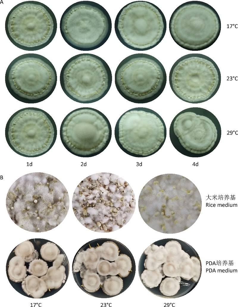
在大型真菌原基形成和子实体发育的过程中,温度是一个极其关键的因素,高温显著影响多种食用菌原基的形成和子实体的品质。广东虫草是中国华南地区特有的虫草类新食品原料,其子实体富含多种活性成分和营养成分,且能够进行较大规模人工栽培。然而,温度对广东虫草原基形成的影响及调控机制尚不清楚。本研究通过不同温度处理不同时间段,发现29℃处理3d抑制了广东虫草原基的形成;对29℃处理3d处理前后的菌丝阶段(CK)和原基阶段(CK-P和HT-P)样品进行转录组测序分析,高温处理后原基阶段的两个组中发现1 682个差异表达基因,其中1 015个上调表达和667个下调表达。在碳水化合物代谢途径中,多个糖酵解和三羧酸循环及葡聚糖和海藻糖合成相关基因在高温处理后呈现下调表达;在原基样品中,多个热激蛋白基因(Hsp10Hsp23DnaJHsp70Hsp90Hsp98)和转录因子C2H2转录水平显著上调表达。本研究结果基于分子水平揭示了高温影响原基形成过程中能量代谢和相关基因的差异表达,为后续利用广东虫草抗逆相关基因资源培育新品种奠定了重要的基础。

Abstract

During the process of primordial formation and fruiting body (FB) development in macro-fungi, temperature is an extremely crucial factor, and high temperature significantly affects primordial formation and FB quality. The fruiting bodies of Cordyceps guangdongensis, a new and special food resource of Cordyceps-like fungi in South China, have abundant bioactive and nutritional ingredients, and are cultivatable in a large scale. Nevertheless, the effect and regulatory mechanisms of temperature on its primordial formation remain less-known. In this study, we found that 3d treatment under 29°C obviously inhibited the primordial formation of T. guangdongense using different period treatments of different temperatures. After treatment, the samples of mycelium (CK) and primordium (CK-P and HT-P) stages were used for transcriptome sequencing. In the comparison of HT-P vs CK-P, 1 682 differentially expressed genes (DEGs) were gained. Among them, 1 015 genes were markedly up-regulated, and 667 genes were obviously down-regulated. In terms of the carbohydrate metabolic pathway, several genes involved in glycolysis, TCA cycle, glucan and trehalose synthesis were significantly down-regulated. In additions, many genes encoding heat shock protein (Hsp10, Hsp23, DnaJ, Hsp70, Hsp90 and Hsp98) and transcription factor C2H2 were markedly up-regulated in primordia. At molecular level, the results of this study revealed the differential expression of energy metabolism and the relative genes during the process of high temperature affecting primordial formation, laying an important foundation for breeding new varieties of C. guangdongensis using the stress-related genes in the future.

关键词

广东虫草 / 高温胁迫 / 原基形成 / 差异表达基因 / 热激蛋白 / 转录因子

Key words

Cordyceps guangdongensis / high temperature stress / primordial formation / differentially expressed genes / heat shock protein / transcription factor

引用本文

导出引用
王刚正, 李敏, 张成花, 孙程远, 李泰辉, 邓旺秋. 基于转录组分析高温抑制广东虫草原基形成的调控网络[J]. 菌物学报, 2021, 40(11): 2940-2952 https://doi.org/10.13346/j.mycosystema.210289
WANG Gang-Zheng, LI Min, ZHANG Cheng-Hua, SUN Cheng-Yuan, LI Tai-Hui, DENG Wang-Qiu. Dissecting the regulation network of high temperature inhibiting primordial formation of Cordyceps guangdongensis based on transcriptomic data[J]. Mycosystema, 2021, 40(11): 2940-2952 https://doi.org/10.13346/j.mycosystema.210289
丛赤壳科Nectriaceae成立于1865年,模式属为丛赤壳属Nectria (Fr.) Fr.。Rossman et al. (1999)根据形态学特征,将广义的丛赤壳类真菌划分为丛赤壳科和生赤壳科Bionectriaceae。丛赤壳科的主要特征包括子座发达或具基部子座,子囊壳肉质,具丛赤壳型中心体,单生至聚生,表生,近球形、球形、倒梨形至椭圆球形,子囊壳颜色鲜艳,KOH+,LA+,子囊壳表面光滑、具疣状物或毛状物,壳壁厚度通常大于25 μm, 子囊圆柱形至柱棒状, 子囊孢子椭圆形至拟纺锤形, 无分隔至具多个分隔,表面平滑、具条纹、小刺或疣状突起,无色至淡黄褐色(Rossman et al. 1999;庄文颖 2013;Lombard et al. 2015)。目前丛赤壳科已知约55属900余种(Lombard et al. 2015),我国累计报道16属100余种(庄文颖 2013; Zeng & Zhuang 2014, 2015, 2016a, 2016b, 2016c, 2017, 2018, 2019, 2020, 2021a, 2021b; Zeng et al. 2018)。 该科真菌主要分布于温带和热带地区,物种多样性丰富,对农林业发展有重要影响,开展资源调查和系统分类研究,将更新对我国种质资源的认识。

1 材料与方法

研究材料主要采自安徽、河南、湖北、云南和西藏等地的自然保护区和森林公园。采用常规研究方法(Rossman et al. 1999),记录子囊壳在3%氢氧化钾(potassium hydroxide,KOH)水溶液和100%乳酸(lactic acid,LA)溶液中的颜色变化。为观察解剖结构特征,将子囊壳置于冷冻切片机YD-1508A(中国金华)上制作厚度约6-8 μm的切片,在解剖镜Olympus SZX7下选取结构完整的切片用乳酚棉兰染色,显微观察其壳壁结构和附属物特征。挑取单个子囊壳制作压片,经乳酚棉兰染色,显微观察子囊和子囊孢子的形状、大小,孢子的颜色、表面纹饰和分隔情况;采用Zeiss Axioskop 2 plus (哥廷根)光学显微镜配备的Canon G5摄像系统拍照。观察菌株在CMD (cornmeal dextrose agar)、PDA (potato dextrose agar)和SNA (synthetic nutrient-poor agar) (Nirenberg 1976)培养基上25 ℃培养7 d的菌落形态,测量菌落直径。
研究标本存放于中国科学院微生物研究所菌物标本馆(herbarium mycologicum academiae sinicae,HMAS),菌种保藏于微生物研究所真菌学国家重点实验室。参照Wang & Zhuang (2004)的方法提取菌丝DNA,使用引物ITS5/ITS4 (White et al. 1990)和LR0R/LR5 (Rehner & Samuels 1994)扩增ITS和LSU序列,获得序列提交至GenBank,使用BioEdit 7.0.5.3 (Hall 1999)进行序列拼接、比对和编辑,运用BLASTN在NCBI (https://www.ncbi.nlm.nih.gov/)数据库进行检索。
本研究综合形态解剖、培养性状、DNA序列和无性阶段等特征,对各标本进行系统分类鉴定。采用最大简约(maximum parsimony,MP)和贝叶斯(Bayesian inference,BI)方法明确其系统发育位置,选取ITS和LSU序列构建系统发育树。进化树中,最大简约分析支持率(bootstrap proportion,BP)大于50%和贝叶斯分析后验概率(posterior probability,PP)大于90%分别显示在各分支节点上。

2 分类

肯达拉赤壳 图1
Cosmospora khandalensis (Thirum. & Sukapure) Gräfenhan & Seifert [as 'khandalense'], in Gräfenhan, Schroers, Nirenberg & Seifert, Stud. Mycol. 68: 96, 2011. Fig. 1
Cephalosporium khandalense Thirum. & Sukapure, in Sukapure & Thirumalachar, Mycologia 58(3): 359, 1966.
图1 肯达拉赤壳 (HMAS 247850)

A-C:25 ℃培养7 d的菌落形态 (A:PDA;B:CMD;C:SNA);D-L:分生孢子梗和分生孢子. 标尺:D-L=10 μm

Fig. 1 Cosmospora khandalensis (HMAS 247850).

A-C: Colonies after 7 d at 25 °C (A: PDA; B: CMD; C: SNA); D-L: Conidiophores and conidia. Bars: D-L=10 μm.

Full size|PPT slide

在PDA培养基上,25 ℃生长7 d菌落直径22-23 mm,表面絮状,气生菌丝致密,白色,产生黄绿色色素;在CMD培养基上,25 ℃生长7 d菌落直径23-24 mm,表面绒毛状,气生菌丝稀疏,白色,产生黄绿色色素;在SNA培养基上,25 ℃生长7 d菌落直径21-23 mm,表面绒毛状,气生菌丝稀疏,白色,产生淡黄绿色色素。无性阶段acremonium型,分生孢子梗无色,不分枝或简单分枝,产孢细胞为单瓶梗,圆柱形,长34-64 μm,基部宽1.5-2.5 μm,顶部宽1.0-1.5 μm;分生孢子卵圆形至椭圆形,末端钝圆,无分隔,无色,表面平滑,2.5-5×1.5-2 μm,末端具黏性,通常聚集成团。
标本:湖北神农架木城哨卡,枯枝上生,2014 Ⅸ 22,郑焕娣、曾昭清、秦文韬、陈凯 10045,HMAS 247850 (ITS、LSU GenBank登录号:OK103798、OK103806)。
世界分布:中国、印度、日本、阿根廷、巴西。
讨论:湖北菌株分离自枯枝,其菌落形态、分生孢子等特征与Sukapure & Thirumalachar (1966)和Herrera et al. (2015)的描述一致。序列分析显示中国材料与产于印度的模式菌株(CBS 356.65) ITS序列仅相差1 bp (522/523),LSU完全相同(796/796)。
翠绿赤壳 图2
Cosmospora viridescens (C. Booth) Gräfenhan & Seifert, in Gräfenhan, Schroers, Nirenberg & Seifert, Stud. Mycol. 68: 96, 2011. Fig. 2
Nectria viridescens C. Booth, Mycol. Papers 73: 89, 1959.
图2 翠绿赤壳 (HMAS 247851)

A-C:25 ℃培养7 d的菌落形态 (A:PDA;B:CMD;C:SNA);D-L:分生孢子梗和分生孢子. 标尺:D-L=10 μm

Fig. 2 Cosmospora viridescens (HMAS 247851).

A-C: Colonies after 7 d at 25 °C (A: PDA; B: CMD; C: SNA); D-L: Conidiophores and conidia. Bars: D-L=10 μm.

Full size|PPT slide

在PDA培养基上,25 ℃生长7 d菌落直径23-24 mm,表面絮状,气生菌丝致密,白色,产生黄色至黄绿色色素;在CMD培养基上,25 ℃生长7 d菌落直径25-26 mm,表面絮状,气生菌丝较稀疏,白色,产生黄绿色色素;在SNA培养基上,25 ℃生长7 d菌落直径25-27 mm,表面绒毛状,气生菌丝稀疏,白色。无性阶段acremonium型,分生孢子梗无色,不分枝或简单分枝,产孢细胞为单瓶梗,圆柱形,长30-68 μm,基部宽1.8-2.5 μm,顶部宽1.0-1.2 μm;分生孢子椭圆形至杆形,末端钝圆,无分隔,无色,表面平滑,3-5×2-3 μm,末端具黏性,少数聚集成团。
标本:西藏米林南伊沟,Ganoderma sp.上生,2016 Ⅸ 13,郑焕娣、曾昭清、王新存、陈凯、张玉博 10806,HMAS 247851 (ITS、LSU GenBank登录号:OK103799、OK103807)。
世界分布:中国、捷克、丹麦、英国。
讨论:西藏菌株的形态特征与Booth (1959)提供的原始描述一致。Gräfenhan et al. (2011)对其形态相近种进行了详细讨论。我国菌株与捷克菌株(CBS 102430)的ITS和LSU序列分别相差2 bp (518/520)和3 bp (785/788),与来自英国的模式菌株(IMI 73377a)相差5 bp (534/539)和6 bp (782/788),将上述差异视为种内变异。这是该种首次在亚洲发现(Herrera et al. 2015)。
剑孢新赤壳 图3
Neocosmospora protoensiformis Sand.-Den. & Crous, in Sandoval-Denis, Lombard & Crous, Persoonia 43: 156, 2019. Fig. 3
Fusarium protoensiforme (Sand.-Den. & Crous) O’Donnell, Geiser, Kasson & T. Aoki, in Aoki, Geiser, Kasson & O'Donnell, Index Fungorum 440: 3, 2020.
图3 剑孢新赤壳 (HMAS 290889)

A-C:自然基物上的子囊壳;D,E:25 ℃培养7 d的菌落形态 (D:PDA;E:SNA);F:子囊壳纵切面结构;G-I:子囊及子囊孢子;J-L:子囊孢子;M,N:分生孢子梗和小型分生孢子;O:小型分生孢子;P-S:大型分生孢子. 标尺:A-C=1 mm;F=50 μm;G-S=10 μm

Fig. 3 Neocosmospora protoensiformis (HMAS 290889).

A-C: Ascomata on natural substratum; D, E: Colonies after 7 d at 25 °C (D: PDA; E: SNA); F: Median section of an ascoma; G-I: Asci with ascospores; J-L: Ascospores; M, N: Conidiophores and microconidia; O: Microconidia; P-S: Macroconidia. Bars: A-C=1 mm; F=50 μm; G-S=10 μm.

Full size|PPT slide

无子座;子囊壳单生至群生,表生,球形至梨形,表面具疣状物,乳突较小,干后侧面明显凹陷,新鲜时为鲜红色,干后为深红色,在3% KOH水溶液中呈暗红色,100%乳酸溶液中呈黄色,高274-363 μm,直径216-294 μm;疣状物高4-40 μm,细胞球形至近球形,8-22×6-20 μm;壳壁厚20-50 μm,细胞矩胞组织至角胞组织,5.4-15×2.2-8 μm,胞壁厚1.0-1.5 μm;子囊棒状,顶部简单,无顶环,具8个孢子,43-60×5-10 μm;子囊孢子椭圆形,具1个分隔,分隔处稍缢缩,无色,表面平滑,在子囊中斜向单列排列,10-15× 5-8 μm。
在PDA培养基上,25℃培养7 d菌落直径40 mm,气生菌丝致密,白色;在SNA培养基上,25 ℃培养7 d菌落直径45 mm,白色,气生菌丝稀疏;分生孢子梗简单分枝,锥形、近圆柱形至针形,表面光滑,长22-56 μm,基部宽2-3 μm,顶部宽1-1.5 μm;大型分生孢子镰刀形,通常一端带小弯钩,具4-9个分隔,50-85×4-5 μm;小型分生孢子卵圆形、棒状至椭圆形,不弯曲,具0(-1)个分隔,无色,表面平滑,8-17(-20)×3-5 μm,末端具黏性,少数聚集成团。
标本:云南高黎贡山百花岭,枯树皮上生,2017 Ⅸ 15,张意、郑焕娣、王新存、张玉博 11363,HMAS 290889 (ITS、LSU GenBank登录号:OK103800、OK103808)。
世界分布:中国、委内瑞拉。
讨论:该种可在人工培养基上产生子囊壳,与自然基物上的相比,子囊壳和子囊孢子的大小基本一致,子囊稍大(53-105×8-13.8 μm vs. 43-60× 5-10 μm) (Sandoval-Denis et al. 2019),我国菌株与产自委内瑞拉的模式菌株(NRRL 22178)的ITS序列相差5 bp (518/523),LSU序列完全一致(535/535)。本研究将上述差异处理为种内变异。
罗杰森假赤壳 图4
Pseudocosmospora rogersonii C.S. Herrera & P. Chaverri, Mycologia 105(5): 1299, 2013. Fig. 4
图4 罗杰森假赤壳 (HMAS 247852)

A,B:25 ℃培养14 d的菌落形态 (A:PDA;B:SNA);C-I:分生孢子梗和分生孢子;J,K:分生孢子. 标尺:C-K=10 μm

Fig. 4 Pseudocosmospora rogersonii (HMAS 247852).

A, B: Colonies after 14 d at 25 °C (A: PDA; B: SNA); C-I: Conidiophores and conidia; J, K: Conidia. Bars: C-K=10 μm.

Full size|PPT slide

在PDA培养基上,25 ℃生长14 d菌落直径37 mm,具壳状,粉红至米褐色,背面同色;在SNA培养基上,25 ℃生长14 d菌落直径15 mm,气生菌丝极稀疏,淡粉色。无性阶段acremonium型,分生孢子梗简单,不分枝,圆柱形,朝顶部渐细,无色,长28-95 μm,基部宽1.2-1.5 μm,顶部宽0.8-1 μm。分生孢子矩形、椭圆形至杆状,不分隔,无色,表面平滑,2.5-5×1-1.8 μm。
标本:安徽金寨天堂寨,真菌上生,2011 Ⅷ 24,陈双林、庄文颖、曾昭清、郑焕娣7889,HMAS 247852 (ITS、LSU GenBank登录号:OK103796、OK103804)。
世界分布:中国、美国。
讨论:与Herrera et al. (2013)基于美国材料对该种的描述相比,我国安徽菌株的分生孢子略小(2.5-5×1-1.8 μm vs. 2.9-5.5×1.1-2.6 μm),其他特征相同。菌株7889的ITS (520/520)和LSU (764/764)序列与模式菌株BPI 1107121完全一致。该种在我国发现使其分布范围由北美洲扩展至亚洲。
瘤顶赤壳 图5
Tumenectria laetidisca (Rossman) Salgado & Rossman, in Salgado-Salazar, Rossman & Chaverri, Fungal Diversity 80: 451, 2016. Fig. 5
Nectria laetidisca Rossman, Mycol. Pap. 150: 36, 1983.
=Cylindrocarpon bambusicola Matsush., Matsush. Mycol. Mem. 5: 9. 1987.
图5 瘤顶赤壳 (HMAS 290890)

A-C:自然基物上的子囊壳;D,E:25 ℃培养14 d的菌落形态 (D:PDA;E:SNA);F:子囊壳纵切面结构;G-K:分生孢子梗和分生孢子;L:厚垣孢子. 标尺:A-C=1 mm;F=50 μm;G-L=10 μm

Fig. 5 Tumenectria laetidisca (HMAS 290890).

A-C: Ascomata on natural substratum; D, E: Colonies after 14 d at 25 °C (D: PDA; E: SNA); F: Median section of an ascoma; G-K: Conidiophores and conidia; L: Chlamydospores. Bars: A-C=1 mm; F=50 μm; G-L=10 μm.

Full size|PPT slide

无子座;子囊壳单生,表生,球形至近球形,顶部具乳突,高38-75 μm,基部宽50-100 μm,顶部宽30-50 μm,干后不凹陷,新鲜时为鲜红色,干后为深红色,在3% KOH水溶液中呈暗红色,100%乳酸溶液中呈黄色,高225-304 μm,直径206-225 μm;壳壁厚28-48 μm,分2层,外层厚23-41 μm,细胞角胞组织至球胞组织,5-13× 3-8 μm,胞壁厚0.8-1.0 μm;内层厚5-7 μm,细胞矩胞组织,8-15×2.5-3.5 μm,胞壁厚0.6-0.8 μm;子囊和子囊孢子未见。
在PDA培养基上,25 ℃生长14 d菌落直径36 mm,表面絮状,气生菌丝致密,白色,背面产生米黄色至淡黄褐色色素;在SNA培养基上,25 ℃生长14 d菌落直径42 mm,表面绒毛状,气生菌丝稀疏,白色。无性阶段cylindrocarpon型,分生孢子梗无色,产孢细胞圆柱形,18-35×3.5- 5 μm;大型分生孢子圆柱形至纺锤形,中间宽,两端略圆,具3-5个分隔,48-77.1×7.4-10.9 μm;偶见厚垣孢子,球形至近球形,直径5-18 μm,间生或串生。
标本:河南洛阳重渡沟,枯枝上生,2013 Ⅸ 20,郑焕娣、曾昭清、朱兆香8813,HMAS 290890 (ITS、LSU GenBank登录号:OK103797、OK103805)。
世界分布:中国、日本、牙买加。
讨论:该种曾被纳入Nectria,综合形态学特征和分子系统学证据,Salgado-Salazar et al. (2016)以其为模式种建立新属Tumenectria Salgado & Rossman,目前仅包括1个种。河南材料状态不佳,子囊壳数量很少,未观察到子囊和子囊孢子,其无性阶段特征符合Salgado-Salazar et al. (2016)的描述。中国菌株(8813)与日本菌株(CBS 100284)的ITS (478/478)和LSU (797/797)序列完全一致,而与牙买加的模式菌株(CBS 101909)分别相差5 bp (473/478)和0 bp (797/797)。

3 系统发育分析

为了清晰地显示5个中国新记录种的系统发育位置,选择丛赤壳科的7个种14个菌株的ITS和LSU序列,以Stachybotrys chartarum为外群,运用MP和BI方法分别构建系统发育树。结果显示,BI树和MP树的拓扑结构一致,最大简约分析产生的唯一进化树(图6)显示菌株HMAS 247850、247851、290889、247852和290890分别与Cosmospora khandalensisCosmospora viridescensNeocosmospora protoensiformisPseudocosmospora rogersoniiTumenectria laetidisca聚类在一起,从而支持了上述形态学研究的结果。
图6 基于ITS和LSU序列的MP树

粗体显示5个中国新记录种的系统发育位置,MPBP大于50% (左)、BIPP大于90% (右)标注于分支节点上

Fig. 6 Maximum parsimony phylogram reconstructed from the combined sequences of ITS and LSU.

the phylogenetic position of the five Nectriaceae species new to China. MPBP above 50% (left) showing and BIPP above 90% (right) are given respectively.

Full size|PPT slide

参考文献

[1]
Cao XT, Bian YB, Xiao XJ, Li JS, Wang GZ, 2015. Effect of heat stress on Lentinula edodes mycelial growth recovery and resistance to Trichoderma harzianum. Acta Edulis Fungi, 22(4):81-85 (in Chinese)
[2]
Cui HY, Li TH, Song B, Yan WJ, 2010. GC-MS analysis of fatty acids from Cordyceps guangdongensis. Acta Edulis Fungi, 17(2):89-92 (in Chinese)
[3]
Dai YC, Yang ZL, 2008. A revised checklist of medicinal fungi in China. Mycosystema, 27:801-824 (in Chinese)
[4]
Kim D, Langmead B, Salzberg S, 2015. HISAT: a fast spliced aligner with low memory requirements. Nature Methods, 12(4):357-360
Kim, Daehwan; Langmead, Ben; Salzberg, Steven L. Johns Hopkins Univ, Sch Med, Ctr Computat Biol, McKusick Nathans Inst Genet Med, Baltimore, MD 21205 USA. Kim, Daehwan Johns Hopkins Univ, Dept Biostat, Bloomberg Sch Publ Hlth, Baltimore, MD 21205 USA. Langmead, Ben; Salzberg, Steven L. Johns Hopkins Univ, Dept Comp Sci, Baltimore, MD 21218 USA.
[5]
Lechner BE, Alberto E, 2007. Optimal conditions for the fruit body production of natural occurring strains of Lentinus tigrinus. Bioresource Technology, 98(9):1866-1869
[6]
Lin QY, Li TH, Huang H, Song B, 2009a. Studies on light and temperature conditions for cultivation of Cordyceps guangdongensis. Journal of South China Agricultural University, 30(1):42-45 (in Chinese)
[7]
Lin QY, Li TH, Song B, Huang H, 2009b. Comparison of selected chemical component levels in Cordyceps guangdongensis, C. sinensis and C. militaris. Acta Edulis Fungi, 16(4):54-57 (in Chinese)
[8]
Liu L, Yang C, Luo Q, Xu N, Zhou MZ, Gao B, Wang C, Shi Y, 2018. Fermenting liquid vinegar with higher taste, flavor and healthy value by using discarded Cordyceps militaris solid culture medium. LWT-Food Science and Technology, 98:654-660
[9]
Liu XM, Huang CY, Chen Q, Wu XL, Zhang JX, 2013. Alleviative effects of exogenous trehalose on oxidative damage metabolism in Pleurotus pulmonarius under heat stress. Acta Horticulturae Sinica, 40(8):1501-1508 (in Chinese)
[10]
Love MI, Huber W, Anders S, 2014. Moderated estimation of fold change and dispersion for RNA-seq data with DESeq2. Genome Biology, 15(12):550
[11]
Lu Z, Kong X, Lu Z, Xiao MX, Chen MY, Zhu L, Shen YM, Hu XY, Song SY, 2014. Para-aminobenzoic acid (paba) synthase enhances thermotolerance of mushroom Agaricus bisporus. PLoS One, 9(3):e91298
[12]
Manzi P, Pizzoferrato L, 2000. Beta-glucans in edible mushrooms. Food Chemistry, 68(3):315-318
[13]
Meng C, Han Q, Wang X, Liu XC, Fan XY, Liu RN, Wang Q, Wang CY, 2019. Determination and quantitative comparison of nucleosides in two Cordyceps by HPLC-ESI-MS-MS. Journal of Chromatographic Science, 57(5):426-433
[14]
Ohm RA, de Jong JF, de Bekker C, Wösten HAB, Lugones LG, 2011. Transcription factor genes of Schizophyllum commune involved in regulation of mushroom formation. Molecular Microbiology, 81:1433-1445
[15]
Pelkmans JF, Vos AM, Scholtmeijer K, Hendrix E, Baars JJP, Gehrmann T, Reinders MJT, Lugones LG, Wösten HAB, 2016. The transcriptional regulator c2h2 accelerates mushroom formation in Agaricus bisporus. Applied Microbiology and Biotechnology, 100(16):7151-7159
The Cys2His2 zinc finger protein gene c2h2 of Schizophyllum commune is involved in mushroom formation. Its inactivation results in a strain that is arrested at the stage of aggregate formation. In this study, the c2h2 orthologue of Agaricus bisporus was over-expressed in this white button mushroom forming basidiomycete using Agrobacterium-mediated transformation. Morphology, cap expansion rate, and total number and biomass of mushrooms were not affected by over-expression of c2h2. However, yield per day of the c2h2 over-expression strains peaked 1 day earlier. These data and expression analysis indicate that C2H2 impacts timing of mushroom formation at an early stage of development, making its encoding gene a target for breeding of commercial mushroom strains.
[16]
Qiu Z, Wu X, Zhang J, Huang C, 2018. High- temperature induced changes of extracellular metabolites in Pleurotus ostreatus and their positive effects on the growth of Trichoderma asperellum. Frontiers in Microbiology, 9:10
[17]
Tarazona S, García-Alcalde F, Dopazo J, Ferrer A, Conesa A, 2011. Differential expression in RNA-Seq: a matter of depth. Genome Research, 21(12):2213-2223
Next-generation sequencing (NGS) technologies are revolutionizing genome research, and in particular, their application to transcriptomics (RNA-seq) is increasingly being used for gene expression profiling as a replacement for microarrays. However, the properties of RNA-seq data have not been yet fully established, and additional research is needed for understanding how these data respond to differential expression analysis. In this work, we set out to gain insights into the characteristics of RNA-seq data analysis by studying an important parameter of this technology: the sequencing depth. We have analyzed how sequencing depth affects the detection of transcripts and their identification as differentially expressed, looking at aspects such as transcript biotype, length, expression level, and fold-change. We have evaluated different algorithms available for the analysis of RNA-seq and proposed a novel approach--NOISeq--that differs from existing methods in that it is data-adaptive and nonparametric. Our results reveal that most existing methodologies suffer from a strong dependency on sequencing depth for their differential expression calls and that this results in a considerable number of false positives that increases as the number of reads grows. In contrast, our proposed method models the noise distribution from the actual data, can therefore better adapt to the size of the data set, and is more effective in controlling the rate of false discoveries. This work discusses the true potential of RNA-seq for studying regulation at low expression ranges, the noise within RNA-seq data, and the issue of replication.
[18]
Wang GZ, Li M, Zhang CH, Cheng HJ, Gao Y, Deng WQ, Li TH, 2020a. Transcriptome and proteome analyses reveal the regulatory networks and metabolite biosynthesis pathways during the development of Tolypocladium guangdongense. Computational and Structural Biotechnology Journal, 18:2081-2094
[19]
Wang GZ, Li M, Zhang CH, Zhan N, Cheng HJ, Sun CY, Deng WQ, Li TH, 2021. Identification of microRNA-like RNAs in Cordyceps guangdongensis and their expression profile under differential developmental stages. Fungal Genetics and Biology, 147(1):103505
[20]
Wang GZ, Luo Y, Wang C, Zhou Y, Mou CY, Kang H, Xiao Y, Bian YB, Gong YH, 2020b. Hsp40 protein LeDnaJ07 enhances the thermotolerance of Lentinula edodes and regulates IAA biosynthesis by interacting LetrpE. Frontiers in Microbiology, 11:707
[21]
Wang GZ, Zhou SS, Luo Y, Ma CJ, Gong YH, Zhou Y, Gao SS, Huang ZC, Yan LL, Hu Y, Bian YB, 2018. The heat shock protein 40 LeDnaJ regulates stress resistance and indole-3-acetic acid biosynthesis in Lentinula edodes. Fungal Genetics and Biology Journal, 118:37-44
[22]
Wu F, Zhou LW, Yang ZL, Bau T, Li TH, Dai YC, 2019. Resource diversity of Chinese macrofungi: edible, medicinal and poisonous species. Fungal Diversity, 98:1-76
[23]
Xu J, Huang Y, Chen XX, Zheng SC, Chen P, Mo MH, 2016. The mechanisms of pharmacological activities of Ophiocordyceps sinensis fungi. Phytotherapy Research, 30(10):1572-1583
[24]
Yan WJ, Li TH, Lao J, Song B, Shen YH, 2013. Anti-fatigue property of Cordyceps guangdongensis and the underlying mechanisms. Pharmaceutical Biology, 51(5):614-620
[25]
Yan WJ, Li TH, Lin QY, Song B, Jiang ZD, 2010. Safety assessment of Cordyceps guangdongensis, 2010. Food and Chemical Toxicology, 48(11):3080-3084
[26]
Yan WJ, Li TH, Zhong Z, 2014. Anti-inflammatory effect of a novel food Cordyceps guangdongensis on experimental rats with chronic bronchitis induced by tobacco smoking. Food & Function, 5(10):2552-2557
[27]
Yang L, Smyth GK, Wei S, 2014. FeatureCounts: an efficient general purpose program for assigning sequence reads to genomic features. Bioinformatics, 7:923-930
[28]
Zhang CH, Deng WQ, Yan WJ, Li TH, 2018. Whole genome sequence of an edible and potential medicinal fungus, Cordyceps guangdongensis. G3 Genes Genomomes Genetics, 8(6):1863-1870
[29]
曹现涛, 边银丙, 肖新军, 李进山, 王刚正, 2015. 高温胁迫对香菇菌丝生长及其抗哈茨木霉能力的影响. 食用菌学报, 22(4):81-85
[30]
崔红燕, 李泰辉, 宋斌, 闫文娟, 2010. 广东虫草脂肪酸的GC-MS分析. 食用菌学报, 17(2):89-92
[31]
戴玉成, 杨祝良, 2008. 中国药用真菌名录及部分名称的修订. 菌物学报, 27:801-824
[32]
林群英, 李泰辉, 黄浩, 宋斌, 2009a. 广东虫草人工栽培的光温条件研究. 华南农业大学学报, 30(1):42-45
[33]
林群英, 李泰辉, 宋斌, 黄浩, 2009b. 广东虫草与冬虫夏草及蛹虫草的成分比较. 食用菌学报, 16(4):54-57
[34]
刘秀明, 黄晨阳, 陈强, 邬向丽, 张金霞, 2013. 外源海藻糖对高温胁迫下肺形侧耳氧化损伤的缓解效应. 园艺学报, 40(8):1501-1508

基金

广东省科学院专项基金(2020GDASYL-20200103014)
国家自然科学基金(31900015)
广东省科技计划项目(2019B121202005)
广东省科技计划项目(2018B030320001)

版权

版权所有,未经授权,不得转载、摘编本刊文章,不得使用本刊的版式设计。
PDF(1037 KB)

Accesses

Citation

Detail

段落导航
相关文章

/