


灰树花Grifola frondosa (Dicks.) Gray俗称“栗子蘑”,自然条件下主要生长在壳斗科木材上(Dai 2012;吴芳等 2020),特别是栗树根基部,是食、药兼用蕈菌(曹秀明等 2019)。在中医药学里,它具有益气健脾、补虚扶正的功效。主治脾虚气弱、体倦乏力、神疲懒言、饮食减少、食后腹胀,及肿瘤患者放疗或化疗后的上述症状(Wu et al. 2019;Wu et al. 2021)。灰树花中主要的生物活性成分是多糖类物质,灰树花多糖(polysaccharides of Griflola frondosa,PGF)的抗肿瘤、抗癌及免疫调节作用多有报道(郭淑臻 2014)。但国内目前PGF对乳腺癌细胞MCF-7的增殖抑制及作用机制鲜有报道。本研究探讨了不同给药浓度的PGF对MCF-7细胞增殖的抑制作用,给药后MCF-7细胞中Bax/Bcl-2/Caspase-3凋亡通路因子的蛋白表达,旨在为PGF药效学研究提供数据支持,并对灰树花的开发与利用提供参考。
1 材料与方法
1.1 供试材料
1.1.1 试剂
胎牛血清(10099141C),DMEM高糖培养基(C11995500BT),青霉素、链霉素混合液(15140122)、胰蛋白酶-EDTA消化液(25200-072)、10×PBS缓冲液(70013-032)、Invitrogen™ Hoechst 33258水溶液(H3569)购自赛默飞世尔科技有限公司;MTT细胞增殖及细胞毒性检测试剂盒(C0009S)、Annexin V-FITC细胞凋亡检测试剂盒(C1062L)购自碧云天生物技术有限公司;二甲基亚砜(DMSO)购自Amresco公司;Bax (sc-8044)、Bcl-2 (sc-7382)抗体购自Santa Cruz生物技术有限公司;β-Actin (4967)、AKT (9272)、p-AKT (9271)、m-TOR (2972)、p-m-TOR (5536)购自Cell Signaling生物技术有限公司;灰树花购自江中食疗科技有限公司。
1.1.2 仪器
超净工作台(Thermo公司);Synergy LX型酶标仪(BioTek公司);HF180 CO2培养箱(HealForce公司);Countess 3 FL自动细胞计数仪(Invitrogen公司);Guava Muse细胞分析仪(Luminex公司);96孔培养板 (无锡耐思生物科技有限公司);KDC-140HR冷冻离心机(安徽中科中佳科学仪器有限公司);ChemiDoc XRS+IMAGELAB型成像系统(Bio-Rad公司)。
1.2 PGF的提取
称取灰树花粉末,并加入约30倍体积蒸馏水,于70 ℃、500 W超声下辅助提取30 min,95 ℃水浴振荡浸提1 h。离心并取上清液,浓缩后加入4倍体积无水乙醇,4 ℃过夜。沉淀依次经过无水乙醇、丙酮和乙醚洗涤。离心取沉淀物,冷冻干燥后得灰树花粗多糖。将Sevage试剂与糖液按1:3体积混合,振摇30 min,静置分层,取上层多糖溶液,重复多次。将多糖溶液浓缩装入透析袋(截留分子量8 000)中,蒸馏水透析96 h,真空冷冻干燥后得纯化后的多糖,-20 ℃冷冻保存备用。采用硫酸-苯酚法测定PGF提取物多糖含量达90.24%。
1.3 MCF-7细胞株培养
常规方法复苏冻存于液氮罐中的人乳腺癌细胞-7(MCF-7),将MCF-7接种于含10% FBS 的RPMI-1640培养基,培养基中添加100 000 U/L青霉素和0.1 g/L的链霉素,置于培养箱中常规培养(37 ℃、5% CO2饱和湿度),每隔3 d换液,待细胞生长融合达80%-90%时,取对数生长期的细胞进行实验。
1.4 MCF-7细胞抑制实验
MCF-7细胞分为不同PGF浓度给药组、阴性对照组及空白对照组,其中空白对照组只加入等量完全培养基不接种细胞。对数生长期的MCF-7细胞分别按1×10⁴个/孔接种于96孔培养板,200 μL/孔,置37 ℃的5% CO2培养箱培养,待细胞贴壁后,弃去旧培养基,加入含不同浓度PGF (1、10、20、50、100和200 μg/mL)的培养基200 μL,空白对照组和阴性对照组加入不含PGF的完全培养基200 μL。将MCF-7细胞继续培养24、48和72 h后弃去上清液,每孔加入5 mg/mL的MTT溶液15 μL,继续培养4 h,取出各组细胞,弃上清液后各孔加入DMSO 150 μL,置摇床振荡20 min;酶标仪设定波长为450 nm,记录各组细胞相应吸光度值(A值)。根据A值计算PGF对5种肿瘤细胞的抑制率,以各肿瘤细胞空白对照组作为调零孔,A实验组= A不同浓度的PGF给药组-A空白对照组,A对照组=A阴性对照组-A空白对照组,抑制率(%)=(1-A不同浓度的PGF给药组/A对照组)×100%。每组设3个复孔。
1.5 Hoechst染色
取对数期生长的MCF-7细胞,按4×10⁵个/孔接种于Thermanox塑料盖片上,37 ℃、5% CO2条件下培养。实验分为空白对照组(Nor)、低剂量组(PGF20)、中剂量组(PGF50)以及高剂量组(PGF100)。细胞汇合至50%时,给药继续培养24 h,给药后使用Hoechst33258试剂盒按说明书操作指南进行染色。中性树脂封片,通过荧光显微镜观察细胞形态。
1.6 细胞凋亡的流式检测
MCF-7细胞分为对照组与不同PGF浓度给药组。对数生长期的MCF-7细胞按1×10⁵个/孔分别接种于6孔培养板,置37 ℃的5% CO2培养箱培养,待细胞贴壁后,弃去旧培养基,加入含不同浓度PGF (20、50和100 μg/mL)的培养基3 mL,对照组加入不含PGF的完全培养基3 mL。将MCF-7细胞继续培养24 h后弃去上清液,加1 mL预冷处理的1×PBS清洗2次,细胞消化后1 000 r/mim离心5 min后,收集细胞沉淀。用400 μL的1×Annexin V 结合液重悬细胞,混合均匀,细胞浓度控制在1×10⁶以内。向细胞悬液中加5 μL Annexin V-FITC染色剂,混匀后4 ℃培养15 min。混合液中最后加入10 μL PI染色剂,混匀后4℃避光培养15 min。孵育后立即用流式细胞仪进行检测。
1.7 Western blotting
不同浓度(20、50和100 μg/mL)的PGF给药24 h后,收获MCF-7细胞,用100 μL RIPA裂解液涡旋裂解细胞,4 ℃、14 000 r/min离心20 min,上清液即为细胞总蛋白液。BCA法检测总蛋白浓度。蛋白采用十二烷基硫酸钠-聚丙烯酰胺凝胶电泳(SDS-PAGE)进行分离,聚偏二氟乙烯膜(PVDF) 4 ℃进行转膜2 h。5%脱脂奶粉室温封闭2 h。一抗Bax (1:300)、Bcl-2 (1:500)、β-Actin (1:2 000)、AKT (1:500)、p-AKT (1:500)、m-TOR (1:1 000)、p-m-TOR (1:1 000) 4 ℃孵育过夜,洗膜缓冲液(TBST)清洗3次、每次5 min,二抗anti-mouse (1:2 000)、anti-rabbit (1:2 000)室温孵育2 h后TBST再次清洗3次、每次5 min。最后ECL显色法于Bio-Rad成像系统下显影,以β-Actin为内参,计算相关蛋白表达水平。
1.8 统计分析
计量结果以¯x±s表示,细胞活性的抑制对比分析采用SPSS 23.0统计软件进行two-way ANOVA分析,其他数据对比采用one-way ANOVA分析,采用GraphPad Prism 9 (GraphPad Software Inc.)软件绘制相关柱状图。P<0.05时有显著性差异。
2 结果与分析
2.1 PGF对MCF-7细胞活性的抑制作用
为了探讨PGF对MCF-7细胞活性的抑制作用,对MCF-7细胞的增殖抑制率进行检测,不同浓度的PGF对乳腺癌细胞MCF-7分别给药24、48及72 h后的增殖抑制率影响见图1。PGF作用24 h后,20-200 μg/mL PGF对MCF-7细胞增殖抑制率均存在显著性差异(P<0.05)并呈剂量依赖性上升。PGF作用48、72 h后,10-200 μg/mL PGF对MCF-7细胞均存在显著性差异(P<0.05)并呈剂量依赖性上升。实验结果表明,MCF-7细胞给药24 h后20、50及100 μg/mL PGF对细胞增殖抑制率即已呈计量依赖性增高。因此在本研究后续实验中,选取PGF低剂量20 μg/mL、中计量50 μg/mL以及高剂量100 μg/mL为给用浓度,24 h为给药时间。
图1
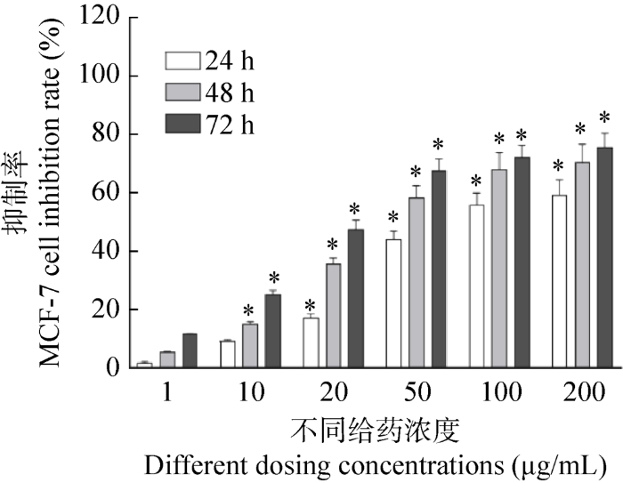
图1
PGF对MCF-7细胞活性的抑制率
*表示不同给药浓度PGF给药24、48及72 h后细胞增殖抑制率对比,P<0.05
Fig. 1
Inhibitory rate of polysaccharides of Grifola frondosa (PGF) on activity of breast cancer cell MCF-7.
* The comparison of cell proliferation inhibition rate after 24, 48 and 72 h treatment of different concentrations of PGF, P<0.05.
2.2 PGF对MCF-7细胞凋亡的影响
为了探讨PGF对MCF-7细胞凋亡的影响,对不同浓度PGF给药后的MCF-7细胞进行了Hoechst染色(图2A),并采用流式细胞检测技术对PGF给药后MCF-7细胞凋亡进行检测 (图2B,2C)。与不同浓度PGF给药24 h后相比,MCF-7正常状态下细胞核形态均一且完整。但PGF 3个给药组与空白组相比,细胞核数量逐渐减少,凋亡小体细胞逐渐增多,即细胞核碎裂现象增多。流式细胞凋亡检测发现,不同浓度的PGF (20、50和100 μg/mL)给药24 h后与对照组相比细胞凋亡总量显著增加,尤其是细胞晚期凋亡数量比例显著增加,并伴随PGF给药浓度的升高呈剂量依赖性增加(图2B)。实验结果表明,PGF呈剂量依赖性促进MCF-7细胞凋亡。
图2
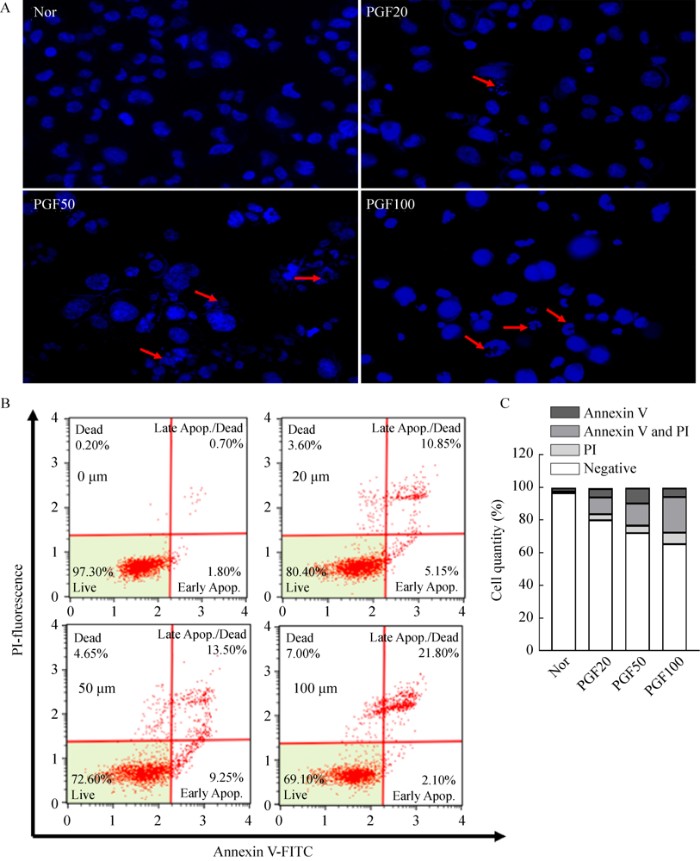
图2
PGF对MCF-7细胞凋亡的影响
A:四组MCF-7细胞Hoechst染色对比;B:流式细胞仪检测细胞凋亡情况;C:细胞数统计图. Negative表示活细胞数,Annexin V阳性表示细胞早期凋亡数,PI阳性表示死亡细胞数量,Annexin V与PI双阳性表示细胞晚期凋亡数量
Fig. 2
Effects of PGF on apoptosis of MCF-7 cells.
A: Comparison of Hoechst staining of MCF-7 cells in four experimental groups; B: Cell apoptosis detected by flow cytometry; C: Statistical diagram of cell number. Negative indicated the number of living cells, Annexin V positive indicated the number of early apoptosis, PI positive indicated the number of dead cells, and Annexin V and PI double positive indicated the number of late apoptosis. Nor: Untreated cell; PGF20: 20 μg/mL; PGF50: 50 μg/mL; PGF100: 100 μg/mL.
2.3 PGF对MCF-7细胞凋亡因子的调节作用
为了探讨PGF对MCF-7细胞凋亡因子的调节作用,采用Western blotting对细胞凋亡调节蛋白Bax、Bcl-2、Pro-Caspase-3和Cleaved- Caspase-3进行表达研究(图3A)。给药24 h后,20、50与100 μg/mL PGF组MCF-7细胞Bax和Cleaved-Caspase-3蛋白表达水平与正常组相比显著升高(P<0.05,图3B,3E);20、50与100 μg/mL PGF组MCF-7细胞Bcl-2、Pro- Caspase-3蛋白表达水平与正常组相比显著降低(P<0.05,图3C,3D)。实验结果表明,PGF可促进MCF-7细胞凋亡从而使MCF-7细胞在给药24 h后Bax、Cleaved-Caspase-3蛋白表达增加、Bcl-2、Pro-Caspase-3蛋白表达减少。
图3
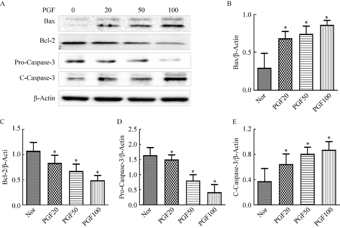
图3
PGF对MCF-7细胞凋亡因子的调节
A:四组MCF-7细胞Western blotting蛋白表达水平;B:Bax蛋白表达水平灰度值统计图;C:Bcl-2蛋白表达水平灰度值统计图;D:Pro-Caspase-3蛋白表达水平灰度值统计图;E:Cleaved-Caspase-3蛋白表达水平灰度值统计图. *给药24 h后PGF不同给药浓度组与对照组的蛋白表达水平相比较差异显著(P<0.05)
Fig. 3
MCF-7 apoptosis factor regulated by PGF.
A: Western blotting protein expression level of MCF-7 cells in four experimental groups; B: Gray scale of Bax protein expression level; C: Gray scale of Bcl-2 protein expression level; D: Statistical figure of the gray level of Pro-Caspase-3 protein expression; E: Gray level graph of Cleaved-Caspase-3 protein expression level. *After 24 h of treatment, the protein expression level of the experimental groups treated with different concentrations of PGF was compared with that of control group (P<0.05). Nor: Untreated cell; PGF20: 20 μg/mL; PGF50: 50 μg/mL; PGF100: 100 μg/mL.
3 讨论
中医认为乳腺癌的发病与肝、肾、脾和胃功能失调有关(陈常莲等 2021),临床研究证明利用中医辨证论治特色,采用疏肝理气、补肾固精或健脾和胃等中医治疗方法,能有效减轻乳腺癌患者术后放疗、化疗的不良作用(吴继萍等 2012),灰树花益气补脾作用显著,在国际上灰树花也被誉为对抗肿瘤的“免疫之王”。近年来相关中药在乳腺癌的应用与疗效机制方面的研究也日渐增多。Bax/Bcl-2/Caspase-3凋亡信号通路是细胞凋亡的关键通路。关于乳腺癌细胞Bax/Bcl-2/Caspase-3细胞凋亡通路,裴岩岩等(2019)研究证明黄芪甲苷可抑制人乳腺癌MCF-7细胞增殖,诱导其凋亡,其作用机制可能与调控此通路有关。但目前灰树花多糖在诱导乳腺癌细胞凋亡及Bax/Bcl-2/Caspase-3细胞凋亡通路机制方面报道较少。
灰树花中主要含有灰树花多糖、甾体、多酚类和维生素类等化学成分(马迪等 2015)。通过对灰树花的现代药理学研究表明,灰树花具有抗肿瘤(刘佳 2018;刘佳等 2019)、抗病毒(高路营 2015)、抗氧化(付佳乐和耿直 2020)、调节免疫力(张婷 2020)和调节血压血脂(刘力萍 2019)等药理作用。中药的多糖类成分例如红花多糖(罗忠兵 2015)、天麻多糖(戴珊珊 2020)和天花粉多糖(曹丽莉等 2012)等在抑制人乳腺癌MCF-7细胞增殖的报道众多。灰树花多糖(PGF)是提取于其菌丝体、子实体或发酵液中的一类含有β-(1-6)、β-(1-6)糖苷键的真菌多糖,具有丰富的中药生物活性(张婷 2020)。近年来国内对PGF进行了大量研究,在肿瘤细胞的研究中,PGF对卵巢癌细胞(吕耀凤和霍颖倩 2012)、肝癌细胞(陶佳等 2019)和肺癌细胞(赵霏 2016)增殖抑制作用均有报道。然而PGF对乳腺癌细胞的增殖抑制作用报道较少。灰树花多糖可分为A、B、C和D不同组分,国外有研究表明灰树花多糖D组分(D组分)可刺激MCF-7细胞凋亡、抑制细胞生长和增殖、阻滞细胞周期、抑制乳腺癌细胞的迁移和转移等作用(Alonso et al. 2013)。但其实灰树花多糖的其他组分均具有一定的抗肿瘤活性,所以本研究对灰树花中的总多糖进行了提取,并对灰树花多糖对乳腺癌MCF-7细胞凋亡的诱导及Bax/Bcl-2/Caspase-3细胞凋亡通路的调节进行了研究。
本研究为探讨PGF抑制乳腺癌细胞增殖的作用机制,采用MTT法测定了PGF不同给药浓度下(1、10、20、50、100和200 μg/mL)以及不同给药时间(24、48、72 h)后MCF-7细胞的增殖抑制率。20、50和100 μg/mL PGF给药24、48和72 h后对MCF-7的增殖均有显著的抑制作用。本实验结果与Martin & Brophy (2010)的研究结果相符,说明PGF抑制乳腺癌细胞增殖呈剂量依赖和时间依赖。为了探讨PGF对MCF-7细胞凋亡的影响,本研究对不同浓度PGF给药24 h后的MCF-7细胞进行可视化荧光染色与流式细胞凋亡检测,结果显示PGF浓度逐渐增加可使MCF-7细胞核数量逐渐减少,细胞核碎裂现象增多,从而诱导细胞凋亡。并且PGF诱导MCF-7细胞晚期凋亡呈剂量依赖性增强。在以上结果的基础上,本研究进一步探讨了PGF对Bax/Bcl-2/Caspase-3细胞凋亡通路相关凋亡因子的蛋白表达的影响。实验采用Western blotting对20、50和100 μg/mL不同浓度下的PGF给药24后MCF-7细胞中Bax、Bcl-2、Pro-Caspase-3和Cleaved-Caspase-3蛋白表达进行检测。研究结果显示20、50和100 μg/mL PGF呈剂量依赖性显著增加MCF-7细胞中Bax、Cleaved-Caspase-3的蛋白表达,同时20、50和100 μg/mL PGF呈剂量依赖性显著减少MCF-7细胞中Bcl-2、Pro-Caspase-3的蛋白表达。细胞凋亡是多基因严格控制的过程(吴军等 2021)。Bax、Bcl-2共属于Bcl-2基因家族。Bcl-2是细胞凋亡抑制基因,Bax不仅有拮抗Bcl-2的抑制凋亡作用,而且具有促进细胞凋亡的功能(刘蕊洁等 2021)。半胱氨酸蛋白酶(caspases)是程序性细胞死亡或凋亡的关键介质。凋亡信号和凋亡受体结合后,作为Caspase family中启动者的Caspase-8和10发生自剪切而被激活后,然后剪切其下游的底物Pro-caspase-3,产生带活性的Cleaved-Caspase-3。本研究对PGF给药后MCF-7细胞中Pro-caspase-3、Cleaved Caspase-3进行检测,结果显示20、50和100 μg/mL的PGF可以显著增加细胞活性 Caspase-3表达水平。研究结果表明PGF可通过凋亡信号激活Bax/Bcl-2/Caspase-3细胞凋亡通路,诱导MCF-7细胞凋亡。
综上所述,本研究证明PGF可呈剂量依赖与时间依赖抑制MCF-7细胞增殖,通过激活Bax/Bcl-2/Caspase-3蛋白通路诱导MCF-7细胞凋亡,为PGF药效学研究提供数据支持,并对灰树花的开发与利用提供参考。
致谢
感谢山东中药大学石俊英教授、李洁教授在Hoechst染色和Western blotting等方面给予的指导;感谢课题组同事们的辛勤付出。
参考文献
Genes related to suppression of malignant phenotype induced by maitake D-fraction in breast cancer cells
DOI:10.1089/jmf.2012.0222
PMID:23875900
[本文引用: 1]

It is already known that the Maitake (D-Fraction) mushroom is involved in stimulating the immune system and activating certain cells that attack cancer, including macrophages, T-cells, and natural killer cells. According to the U.S. National Cancer Institute, polysaccharide complexes present in Maitake mushrooms appear to have significant anticancer activity. However, the exact molecular mechanism of the Maitake antitumoral effect is still unclear. Previously, we have reported that Maitake (D-Fraction) induces apoptosis in breast cancer cells by activation of BCL2-antagonist/killer 1 (BAK1) gene expression. At the present work, we are identifying which genes are responsible for the suppression of the tumoral phenotype mechanism induced by Maitake (D-Fraction) in breast cancer cells. Human breast cancer MCF-7 cells were treated with and without increased concentrations of Maitake D-Fraction (36, 91, 183, 367 μg/mL) for 24 h. Total RNA were isolated and cDNA microarrays were hybridized containing 25,000 human genes. Employing the cDNA microarray analysis, we found that Maitake D-Fraction modified the expression of 4068 genes (2420 were upmodulated and 1648 were downmodulated) in MCF-7 breast cancer cells in a dose-dependent manner during 24 h of treatment. The present data shows that Maitake D-Fraction suppresses the breast tumoral phenotype through a putative molecular mechanism modifying the expression of certain genes (such as IGFBP-7, ITGA2, ICAM3, SOD2, CAV-1, Cul-3, NRF2, Cycline E, ST7, and SPARC) that are involved in apoptosis stimulation, inhibition of cell growth and proliferation, cell cycle arrest, blocking migration and metastasis of tumoral cells, and inducing multidrug sensitivity. Altogether, these results suggest that Maitake D-Fraction could be a potential new target for breast cancer chemoprevention and treatment.
Effects of snakegourd root polysaccharide on apoptosis of MCF-7 cells
Polysaccharides in the fruit body of Grifola frondosa cultivated with JUNCAO
Traditional Chinese medicine zang-fu viscera in the treatment of breast cancer based on syndrome differentiation
Study on extraction, characterization and anti-human breast cancer MCF-7 cells of Gastrodia polysaccharide
Polypore diversity in China with an annotated checklist of Chinese polypores
DOI:10.1007/s10267-011-0134-3 URL [本文引用: 1]
The study of traditional Chinese medicine on cognitive dysfunction of breast cancer patients caused by chemotherapy
Study on antibacterial in vitro and antioxidant activity of Grifola frondosa polysaccharide
Functional polysaccharide of Grifola frondosa inhibit the replication and viability of enterovirus 71
The manufacture technology of Grifola frondosa polysaccharide and its function of falling blood glucose
Study on chemical constituents and pharmacological activities of Grifola frondosa
Antitumor and immune activities of extracts from Grifola frondosa in vivo
Hypoglycemic effects of the exopolysaccharide from submerged culture of Grifola frondosa by addition of rhizoma gastrodiae extracts
Effect of yigan xiaozheng formula on the expression of Bcl-2, Bax mRNA and proteins in rats with preneoplastic liver cells
The effect of safflower polysaccharide on proliferation and apoptosis of breast cancer cell MCF-7
Preliminary investigation on treatment of ovarian cancer by Grifola frondosa polysaccharide in vitro
Chemical components of Grifola frondosa fruiting bodies
Commonly consumed and specialty dietary mushrooms reduce cellular proliferation in MCF-7 human breast cancer cells
DOI:10.1258/ebm.2010.010113 URL [本文引用: 1]
Breast cancer risk prediction models and subsequent tumor characteristics
DOI:10.1007/s12282-020-01060-9
PMID:32056079
[本文引用: 1]
A previous study found evidence that a breast cancer risk prediction model preferentially selected for less aggressive tumors in Swedish women. In the US, the Gail model has been widely used and was used for entry criteria in two large breast cancer prevention trials. We assessed if higher risk levels from the Gail model were associated with less aggressive tumor characteristics and if risk levels were predictive of mortality and survival.We used questionnaire data from women in the Prostate, Lung, Colorectal, and Ovarian Cancer Screening Trial to calculate Gail risk levels (low < 1.66%; moderate 1.66-2.99%; high ≥ 3.00%). Women aged 55-74 were enrolled between 1993 and 2001 and had detailed information on breast cancer incidence and tumors collected. We calculated breast cancer incidence and mortality rates among all women by risk levels and examined breast cancer survival and tumor characteristics among women diagnosed with breast cancer. We used Chi-squared tests and multivariable logistic regression to assess the association between risk levels and tumor characteristics.The study population for this analysis included 45,402 women with 1908 cases of breast cancer. Women at high risk were associated with higher risk of breast cancer mortality compared to women with low risk [rate ratio (RR) = 2.29 95% confidence interval (CI) 1.37-3.84)]. Higher risk levels were associated with lobular-type tumors [moderate: adjusted odds ratio (aOR) = 1.57 95% CI 1.13-2.17; high: aOR = 1.78 95% CI 1.25-2.54] but were not associated with any other tumor characteristics or breast cancer survival.We did not find evidence that higher risk levels from the Gail model are predictive of less aggressive breast cancer tumors.
Astragaloside-Ⅳ induces apoptosis in hunman breast cancer MCF-7 cells via modulating Bax /Bcl-2/Caspase-3 singnaling pathway
Changes in mammographic density over time and the risk of breast cancer: an observational cohort study
DOI:S0960-9776(19)30494-1
PMID:31132476
[本文引用: 1]
The effect of changes in mammographic density over time on the risk of breast cancer remains inconclusive.We used information from four centres of the Breast Cancer Screening Program in Spain in the period 1996-2015. We analysed individual level data from 117,388 women first screened age 50-54, with at least two screening examinations. Breast density was determined using the BI-RADS classification (A to D in increasing order) at earliest and latest screening examination. Adjusted Poisson regression models were used to estimate the relative risk (RR) and 95% confidence intervals (95%CI) of the association between changes in mammographic density and breast cancer risk over time.During an average 5.8 years of follow-up, 1592 (1.36%) women had a breast cancer diagnosis. An increase in density category increased breast cancer risk, and a decrease in density decreased the risk, compared with women who remained in the same BI-RADS category. Women whose density category increased from B to C or B to D had a RR of 1.55 (95%CI = 1.24-1.94) and 2.32 (95%CI = 1.48-3.63), respectively. The RR for women whose density increased from C to D was 1.51 (95%CI = 1.03-2.22). Changes in BI-RADS density were similarly associated with the risk for invasive cancer than for ductal carcinoma in situ.Although a modest proportion of women changed BI-RADS density category, mammographic density changes modulated the risk of breast cancer and identified women at a differential risk. Using two longitudinal measures of BI-RADS density could help target women for risk-based screening strategies.Copyright © 2019 Elsevier Ltd. All rights reserved.
β-glucan from maitake induces lung carcinoma cells apoptosis via activating oxidative stress
Summary of research on tntervention of Chinese medicine in breast cancer treatment
Polypore diversity in South China
Resource diversity of Chinese macrofungi: edible, medicinal and poisonous species
DOI:10.1007/s13225-019-00432-7 URL [本文引用: 1]
Advances in the study of chemotherapy resistance of bcl-2 family in hepatocellular carcinoma
Clinical efficacy evaluation of TCM syndrome differentiation and classification in the treatment of breast cancer
Bioactive ingredients and medicinal values of maitake
Study of Grifola frondosa polysaccharide and vitamin C induced apoptosis and autophagy in hepatocellular carcinoma SMMC-7721 cells
Study of the immunoregulation function of Grifola Frondosa polysaccharide in animal disease model
天花粉多糖诱导人乳腺癌MCF-7细胞凋亡及其Caspase-3和Caspase-8活化对凋亡的影响
益肝消癥方对大鼠肝癌前病变肝细胞凋亡因子Bcl-2、Bax mRNA及蛋白质表达的影响
黄芪甲苷通过Bax/Bcl-2/Caspase-3信号通路诱导人乳腺癌MCF-7细胞凋亡的机制研究


